. 2007 Jul 15;179(2):1282-91.
doi: 10.4049/jimmunol.179.2.1282.

Maternal transmission of resistance to development of allergic airway disease

Affiliations

Maternal transmission of resistance to development of allergic airway disease

Adam P Matson et al. J Immunol. .

Abstract

Parental phenotype is known to influence the inheritance of atopic diseases, such as allergic asthma, with a maternal history being a more significant risk factor for progeny than paternal history. We hypothesized that recall Th1- or Th2-type immune responses during pregnancy would result in transfer of maternal factors that would differentially impact development of immune responsiveness in offspring. Following weaning, susceptibility and severity of allergic airway disease (a murine model of human asthma) was evaluated in progeny, disease being elicited by immunization with OVA-Al(OH)(3) and challenge with aerosolized OVA. We found that progeny of mothers with Th1-biased immunity to OVA subjected to recall aerosol challenge during pregnancy had reduced levels of Ag-specific IgE and airway eosinophilia compared with progeny of mothers with Th2-biased immunity to OVA or naive mothers. Interestingly, progeny of mothers with Th1-type immunity to a heterologous albumin, BSA, were not protected from developing OVA-induced allergic airway disease. These findings demonstrated that maternal transfer of protection from development of allergic airway disease to offspring in this model of maternal Th1-type immunity was Ag specific.


Conflict of interest statement

Disclosures

The authors have no financial conflict of interest.

Figures

FIGURE 1
FIGURE 1
Model to determine the impact of maternal immunity on development of allergic airway disease in offspring. Imm, Immunization (OVA-CFA, OVA-Al(OH)3 or BSA-CFA); Aer, challenge with aerosolized Ag (always matched to immunizing Ag). Parameters of OVA-induced allergic airway disease severity assessed in offspring were OVA-specific Ig levels in serum and composition of leukocytes in the airways.
FIGURE 2
FIGURE 2
Pregnancy does not affect serum levels of OVA-specific IgG1 and IgE. C57BL/6J female mice were immunized three times with OVA-Al(OH)3 (at weekly intervals) and 9 days later were challenged daily, for 7 days, with aerosolized OVA as described in Materials and Methods. Seven weeks later, select females were bred with naive C57BL/6J males and both pregnant and nonpregnant females were re-exposed to aerosolized OVA on days corresponding to E11–17 of pregnancy. Serum was collected 24 h after the last aerosol exposure (E18) and concentrations of OVA-specific IgG1 or IgE in serum were determined by ELISA. Results are from one experiment (three mice per group) and the absence of detectable differences between pregnant and nonpregnant mice was consistent with analyses of eight other mice in separate experiments.
FIGURE 3
FIGURE 3
The profile of OVA-specific cytokine production was determined by the adjuvant used for immunization. C57BL/6J female mice were immunized with OVA-CFA or OVA-Al(OH)3 as described in Materials and Methods. Five days following the second immunization, spleen cells were prepared and OVA-specific cytokine production was determined by ELISPOT. Graphs represent the frequency of cytokine-producing cells per 105 class II-(CD4) or class I-restricted (CD8) T cells, determined from comparisons between T cells cultured in wells containing cognate OVA peptides and control wells (without OVA peptide). Results are expressed as mean ± SE and represent four mice per group analyzed from wells containing three to four dilutions of T cells in duplicate wells. *, p ≤ 0.05 when compared between groups.
FIGURE 4
FIGURE 4
The airway inflammatory response and sensitivity to methacholine observed following aerosol challenge was dependent on the adjuvant used for sensitization. C57BL/6J female mice were immunized with OVA-Al(OH)3 or OVA-CFA and challenged for 10 days with 1% aerosolized OVA (daily exposure time 60 min). A, The differential leukocyte counts in BAL fluid collected 24 h following the last aerosol challenge as determined from analysis of cytocentrifuged slide preparations stained with Wright-Giemsa; B, distribution of T lymphocyte subsets determined by fluorescence flow cytometry; and C, changes in breathing pattern following aerosol challenge with methacholine determined by unrestrained whole body plethysmography in sensitized only (■) or sensitized and challenged mice (▲). Results are expressed as mean ± SE and represent 6 mice per group (A and B) with the exception of the physiology which represent 9–10 mice per group (C). *, p ≤ 0.05 when compared between groups.
FIGURE 5
FIGURE 5
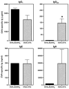
The profiles of maternal OVA-specific Ig isotypes following secondary aerosol challenge during pregnancy were Th1- or Th2-biased dependent on the adjuvant used for sensitization. C57BL/6J female mice were immunized with OVA-Al(OH)3 or OVA-CFA and challenged for 7 days with 1% aerosolized OVA (daily exposure time 60 min). Six weeks later, females were bred with naive C57BL/6J males and pregnant mice were re-exposed to aerosolized OVA on E11–17 of pregnancy. Serum was collected 24 h after the last aerosol exposure (E18) and OVA-specific Igs were measured by ELISA as described in Materials and Methods. Results represent the mean ± SE from four to eight mice per group. Statistical analysis could not be performed for OVA-specific IgE or IgA as analysis of sera was from the OVA-Al(OH)3 mother corresponding to pups shown for IgE and IgA in Fig. 6. *, p ≤ 0.05 when compared between groups.
FIGURE 6
FIGURE 6
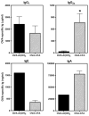
Transmission of Th1- or Th2-biased maternal Abs to offspring was determined by the adjuvant used for immunization. Females were immunized with OVA-Al(OH)3 or OVA-CFA and challenged for 7 days with 1% aerosolized OVA (daily exposure time 60 min). Six weeks later, females were bred with naive C57BL/6J males and pregnant mice were re-exposed to aerosolized OVA on E11–17 of pregnancy. Serum was collected from naive 2-wk-old pups (1 wk before weaning) and concentrations of OVA-specific Igs were measured by ELISA. OVA-specific Igs were absent from serum of pups born to naive C57BL/6J mothers (data not shown). Bar labels refer to conditions of maternal sensitization. Results represent the mean ± SE from three to four determinations per group. Each determination was made using sera from individual mice for IgG1 and IgG2a or sera combined from two mice for IgE and IgA. Statistical analysis could not be performed for OVA-specific IgE or IgA as analysis of sera from progeny of OVA-Al(OH)3 immune mothers was a pooled sample from two mice. *, p ≤ 0.05 when compared between groups.
FIGURE 7
FIGURE 7
Decreased severity of allergic airway disease in offspring of mothers with Th1- but not Th2-type immunity to OVA. Pups were from mothers with Th1- or Th2-type immunity to OVA (sensitized with OVA-CFA or OVA-Al(OH)3 and OVA aerosol challenged 7– 8 wk prior) subjected to secondary challenge with aerosolized OVA during pregnancy. Control pups were from naive mothers never exposed to OVA. Following weaning, allergic airway disease was elicited in 4- to 5-wk-old progeny by two immunizations with OVA-Al(OH)3 followed by challenge for 7 days with 1% aerosolized OVA (daily exposure time 60 min); all pups were immunized and challenged identically. The differences between groups of offspring were restricted solely to factors transmitted as a result of maternal sensitization and exposure to recall OVA during pregnancy. Parameters of disease severity measured in progeny of OVA-immune or naive mothers were (A) OVA-specific IgE levels in serum determined by ELISA; (B) distribution of leukocyte populations in the airways determined from analysis of cytocentrifuged slide preparations stained with Wright-Giemsa; and (C) distribution of T lymphocyte subsets determined by fluorescence flow cytometry. Bar labels refer to conditions of maternal sensitization. Results are expressed as mean ± SE and represent five to eight mice per group. *, p ≤ 0.05 when compared between groups.
FIGURE 8
FIGURE 8
Maternal transmission of resistance to development of allergic airway disease in offspring is Ag specific. Pups were from mothers with Th1-type immunity to OVA or BSA (sensitized with OVA-CFA or BSA-CFA and OVA or BSA aerosol challenged 7– 8 wk prior) subjected to secondary challenge with the respective aerosolized OVA or BSA during pregnancy. Control pups were from naive mothers never exposed to OVA or BSA. Following weaning, allergic airway disease was elicited in 4- to 5-wk-old progeny by two immunizations with OVA-Al(OH)3 followed by challenge for 4 days with 1% aerosolized OVA (daily exposure time 60 min); all pups were immunized and challenged identically. The differences between groups of offspring were restricted solely to factors transmitted as a result of maternal sensitization and exposure to recall Ag during pregnancy. Parameters of disease severity measured in progeny of OVA- or BSA-immune or naive mothers were (A) OVA-specific IgE levels in serum determined by ELISA; (B) distribution of leukocyte populations in the airways determined from analysis of cytocentrifuged slide preparations stained with Wright-Giemsa; and (C) distribution of lymphocytes (CD4+ T cells and CD19+ B cells) determined by fluorescence flow cytometry. Bar labels refer to conditions of maternal sensitization. Results are expressed as mean ± SE and represent five to nine mice per group. *, p ≤ 0.05 when compared with between groups.

Similar articles

Cited by

References

    1. Walker C, Kaegi MK, Braun P, Blaser K. Activated T cells and eosinophilia in bronchoalveolar lavages from subjects with asthma correlated with disease severity. J Allergy Clin Immunol. 1991;88:935–942. - PubMed
    1. McFadden ER, I, Gilbert A. Asthma. N Engl J Med. 1992;327:1928–1924. - PubMed
    1. Corrigan CJ, Kay AB. Asthma: role of T-lymphocytes and lymphokines. Br Med Bull. 1992;48:72– 84. - PubMed
    1. Corrigan CJ, Kay AB. T cells and eosinophils in the pathogenesis of asthma. Immunol Today. 1992;13:501–507. - PubMed
    1. Wilson JW, Djukanovic R, Howarth PH, Holgate ST. Lymphocyte activation in bronchoalveolar lavage and peripheral blood in atopic asthma. Am Rev Respir Dis. 1992;145:958–960. - PubMed

Publication types