

Tumor-associated embryonic antigen-expressing vaccines that target CCR6 elicit potent CD8+ T cell-mediated protective and therapeutic antitumor immunity
- PMID: 17617631
- PMCID: PMC2365706
- DOI: 10.4049/jimmunol.179.2.1381
Tumor-associated embryonic antigen-expressing vaccines that target CCR6 elicit potent CD8+ T cell-mediated protective and therapeutic antitumor immunity
Abstract
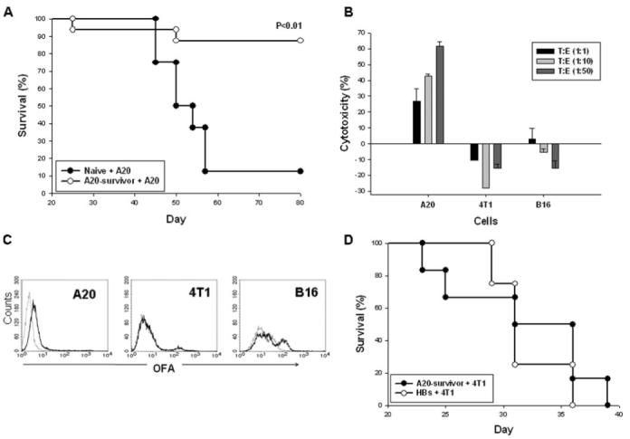
Despite its potency, the wider use of immunotherapy for B cell malignancies is hampered by the lack of well-defined tumor-specific Ags. In this study, we demonstrate that an evolutionarily conserved 37-kDa immature laminin receptor protein (OFA-iLRP), a nonimmunogenic embryonic Ag expressed by a variety of tumors, is rendered immunogenic if targeted to the APCs using the CCR6 ligands MIP3alpha/CCL20 and mDF2beta. The CCR6 targeting facilitated efficient Ag cross-presentation and induction of tumor-neutralizing CTLs. Although the Ag targeting alone, without activation of dendritic cells (DCs), is proposed to induce tolerance, and MIP3alpha does not directly activate DCs, the MIP3alpha-based vaccine efficiently induced protective and therapeutic antitumor responses. The responses were as strong as those elicited by the OFA-iLRP fusions with moieties that activated DCs and Th1-type cytokine responses, mDF2beta, or mycobacterial Hsp70 Ag. Although the same cDNA encodes the dimerized high-affinity mature 67-kDa mLRP that is expressed in normal tissues to stabilize the binding of laminin to cell surface integrins, the vaccines expressing OFA-iLRP elicited long-term protective CD8(+) T cell-mediated memory responses against syngeneic B cell lymphoma, indicating the potential application of these simple vaccines as preventive and therapeutic formulations for human use.
Figures
Similar articles
-
Induction of cytotoxic T-cell responses against the oncofetal antigen-immature laminin receptor for the treatment of hematologic malignancies.Blood. 2003 Dec 15;102(13):4416-23. doi: 10.1182/blood-2003-01-0198. Epub 2003 Jul 17. Blood. 2003. PMID: 12869512
-
Production, safety and antitumor efficacy of recombinant Oncofetal Antigen/immature laminin receptor protein.Biomaterials. 2009 Jun;30(17):3091-9. doi: 10.1016/j.biomaterials.2009.02.022. Epub 2009 Mar 6. Biomaterials. 2009. PMID: 19268360
-
B-cell malignancies as a model for cancer vaccines: from prototype protein to next generation genetic chemokine fusions.Immunol Rev. 1999 Aug;170:115-26. doi: 10.1111/j.1600-065x.1999.tb01333.x. Immunol Rev. 1999. PMID: 10566146 Review.
-
Mediators of innate immunity that target immature, but not mature, dendritic cells induce antitumor immunity when genetically fused with nonimmunogenic tumor antigens.J Immunol. 2001 Dec 1;167(11):6644-53. doi: 10.4049/jimmunol.167.11.6644. J Immunol. 2001. PMID: 11714836
-
CCR6 and CCL20: partners in intestinal immunity and lymphorganogenesis.Ann N Y Acad Sci. 2006 Aug;1072:52-61. doi: 10.1196/annals.1326.036. Ann N Y Acad Sci. 2006. PMID: 17057190 Review.
Cited by
-
Dendritic cell targeted Ccl3- and Xcl1-fusion DNA vaccines differ in induced immune responses and optimal delivery site.Sci Rep. 2019 Feb 12;9(1):1820. doi: 10.1038/s41598-018-38080-7. Sci Rep. 2019. PMID: 30755656 Free PMC article.
-
Chemokines as Cancer Vaccine Adjuvants.Vaccines (Basel). 2013 Dec 1;1(4):444-62. doi: 10.3390/vaccines1040444. Vaccines (Basel). 2013. PMID: 24967094 Free PMC article.
-
MIP-3α-antigen fusion DNA vaccine enhances sex differences in tuberculosis model and alters dendritic cell activity early post vaccination.Sci Rep. 2025 Jul 1;15(1):22264. doi: 10.1038/s41598-025-06532-6. Sci Rep. 2025. PMID: 40596433 Free PMC article.
-
MIP3α-RelMtb intranasal DNA vaccination induces reactive T-cell infiltration into the lungs of mice and macaques.Vaccine. 2025 Jul 16;62:127517. doi: 10.1016/j.vaccine.2025.127517. Online ahead of print. Vaccine. 2025. PMID: 40675108 Free PMC article.
-
Looking into laminin receptor: critical discussion regarding the non-integrin 37/67-kDa laminin receptor/RPSA protein.Biol Rev Camb Philos Soc. 2016 May;91(2):288-310. doi: 10.1111/brv.12170. Epub 2015 Jan 28. Biol Rev Camb Philos Soc. 2016. PMID: 25630983 Free PMC article. Review.
References
-
- Coggin JH, Jr., Barsoum AL, Rohrer JW. 37 kiloDalton oncofetal antigen protein and immature laminin receptor protein are identical, universal T-cell inducing immunogens on primary rodent and human cancers. Anticancer Res. 1999;19:5535–5542. - PubMed
-
- Castronovo V, Claysmith AP, Barker KT, Cioce V, Krutzsch HC, Sobel ME. Biosynthesis of the 67 kDa high affinity laminin receptor. Biochem. Biophys. Res. Commun. 1991;177:177–183. - PubMed
-
- Bendandi M, Gocke CD, Kobrin CB, Benko FA, Sternas LA, Pennington R, Watson TM, Reynolds CW, Gause BL, Duffey PL, et al. Complete molecular remissions induced by patient-specific vaccination plus granulocyte-monocyte colony-stimulating factor against lymphoma. Nat. Med. 1999;5:1171–1177. see comments. - PubMed
-
- Timmerman JM. Immunotherapy for lymphomas. Int. J. Hematol. 2003;77:444–455. - PubMed
Publication types
MeSH terms
Substances
Grants and funding
LinkOut - more resources
Full Text Sources
Other Literature Sources
Research Materials
Miscellaneous
