Impairment of alternative splice sites defining a novel gammaretroviral exon within gag modifies the oncogenic properties of Akv murine leukemia virus
- PMID: 17617899
- PMCID: PMC1936429
- DOI: 10.1186/1742-4690-4-46
Impairment of alternative splice sites defining a novel gammaretroviral exon within gag modifies the oncogenic properties of Akv murine leukemia virus
Abstract
Background: Mutations of an alternative splice donor site located within the gag region has previously been shown to broaden the pathogenic potential of the T-lymphomagenic gammaretrovirus Moloney murine leukemia virus, while the equivalent mutations in the erythroleukemia inducing Friend murine leukemia virus seem to have no influence on the disease-inducing potential of this virus. In the present study we investigate the splice pattern as well as the possible effects of mutating the alternative splice sites on the oncogenic properties of the B-lymphomagenic Akv murine leukemia virus.
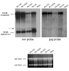
Results: By exon-trapping procedures we have identified a novel gammaretroviral exon, resulting from usage of alternative splice acceptor (SA') and splice donor (SD') sites located in the capsid region of gag of the B-cell lymphomagenic Akv murine leukemia virus. To analyze possible effects in vivo of this novel exon, three different alternative splice site mutant viruses, mutated in either the SA', in the SD', or in both sites, respectively, were constructed and injected into newborn inbred NMRI mice. Most of the infected mice (about 90%) developed hematopoietic neoplasms within 250 days, and histological examination of the tumors showed that the introduced synonymous gag mutations have a significant influence on the phenotype of the induced tumors, changing the distribution of the different types as well as generating tumors of additional specificities such as de novo diffuse large B cell lymphoma (DLBCL) and histiocytic sarcoma. Interestingly, a broader spectrum of diagnoses was made from the two single splice-site mutants than from as well the wild-type as the double splice-site mutant. Both single- and double-spliced transcripts are produced in vivo using the SA' and/or the SD' sites, but the mechanisms underlying the observed effects on oncogenesis remain to be clarified. Likewise, analyses of provirus integration sites in tumor tissues, which identified 111 novel RISs (retroviral integration sites) and 35 novel CISs (common integration sites), did not clearly point to specific target genes or pathways to be associated with specific tumor diagnoses or individual viral mutants.
Conclusion: We present here the first example of a doubly spliced transcript within the group of gammaretroviruses, and we show that mutation of the alternative splice sites that define this novel RNA product change the oncogenic potential of Akv murine leukemia virus.
Figures


Similar articles
-
A 38 nt region and its flanking sequences within gag of Friend murine leukemia virus are crucial for splicing at the correct 5' and 3' splice sites.Microbiol Immunol. 2014 Jan;58(1):38-50. doi: 10.1111/1348-0421.12114. Microbiol Immunol. 2014. PMID: 24236664
-
Activation of c-myb by 5' retrovirus promoter insertion in myeloid neoplasms is dependent upon an intact alternative splice donor site (SD') in gag.Virology. 2004 Dec 20;330(2):398-407. doi: 10.1016/j.virol.2004.09.038. Virology. 2004. PMID: 15567434
-
B-Cell lymphoma induction by akv murine leukemia viruses harboring one or both copies of the tandem repeat in the U3 enhancer.J Virol. 1998 Jul;72(7):5745-56. doi: 10.1128/JVI.72.7.5745-5756.1998. J Virol. 1998. PMID: 9621033 Free PMC article.
-
Host-gene control of C-type tumor virus-expression and tumorigenesis: relevance of studies in inbred mice to cancer in man and other species.Proc Natl Acad Sci U S A. 1971 Nov;68(11):2664-8. doi: 10.1073/pnas.68.11.2664. Proc Natl Acad Sci U S A. 1971. PMID: 4941983 Free PMC article. Review.
-
Alternate forms of myb: consequences of virus insertion in myeloid tumorigenesis and alternative splicing in normal development.Curr Top Microbiol Immunol. 1989;149:71-6. doi: 10.1007/978-3-642-74623-9_6. Curr Top Microbiol Immunol. 1989. PMID: 2659283 Review. No abstract available.
Cited by
-
Protection against retrovirus pathogenesis by SR protein inhibitors.PLoS One. 2009;4(2):e4533. doi: 10.1371/journal.pone.0004533. Epub 2009 Feb 19. PLoS One. 2009. PMID: 19225570 Free PMC article.
-
Characterization of a natural heterodimer between MLV genomic RNA and the SD' retroelement generated by alternative splicing.RNA. 2007 Dec;13(12):2266-76. doi: 10.1261/rna.713807. Epub 2007 Oct 10. RNA. 2007. PMID: 17928575 Free PMC article.
-
Forward and Reverse Genetics of B Cell Malignancies: From Insertional Mutagenesis to CRISPR-Cas.Front Immunol. 2021 Aug 13;12:670280. doi: 10.3389/fimmu.2021.670280. eCollection 2021. Front Immunol. 2021. PMID: 34484175 Free PMC article. Review.
-
Antisense transcription in gammaretroviruses as a mechanism of insertional activation of host genes.J Virol. 2010 Apr;84(8):3780-8. doi: 10.1128/JVI.02088-09. Epub 2010 Feb 3. J Virol. 2010. PMID: 20130045 Free PMC article.
-
A retroviral mutagenesis screen identifies Cd74 as a common insertion site in murine B-lymphomas and reveals the existence of a novel IFNgamma-inducible Cd74 isoform.Mol Cancer. 2010 Apr 23;9:86. doi: 10.1186/1476-4598-9-86. Mol Cancer. 2010. PMID: 20416035 Free PMC article.
References
-
- Rosenberg N., and P. Jolicoeur . Retroviral pathogenesis . In: Coffin JMHSHHEV, editor. Retroviruses. Cold Spring Harbor Laboratory Press, USA; 1997. pp. 475–586. - PubMed
-
- Joosten M, Vankan-Berkhoudt Y, Tas M, Lunghi M, Jenniskens Y, Parganas E, Valk PJ, Lowenberg B, van den Akker E, Delwel R. Large-scale identification of novel potential disease loci in mouse leukemia applying an improved strategy for cloning common virus integration sites. Oncogene. 2002;21:7247–7255. doi: 10.1038/sj.onc.1205813. - DOI - PubMed
Publication types
MeSH terms
Substances
Grants and funding
LinkOut - more resources
Full Text Sources

