

The use of fluorescence enhancement to improve the microscopic diagnosis of falciparum malaria
- PMID: 17617912
- PMCID: PMC1950880
- DOI: 10.1186/1475-2875-6-89
The use of fluorescence enhancement to improve the microscopic diagnosis of falciparum malaria
Abstract
Background: Giemsa staining of thick blood smears remains the "gold standard" for detecting malaria. However, this method is not very good for diagnosing low-level infections. A method for the simultaneous staining of Plasmodium-parasitized culture and blood smears for both bright field and fluorescence was developed and its ability to improve detection efficiency tested.
Methods: A total of 22 nucleic acid-specific fluorescent dyes were tested for their ability to provide easily observable staining of Plasmodium falciparum-parasitized red blood cells following Giemsa staining.
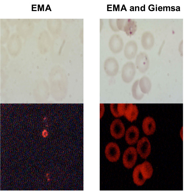
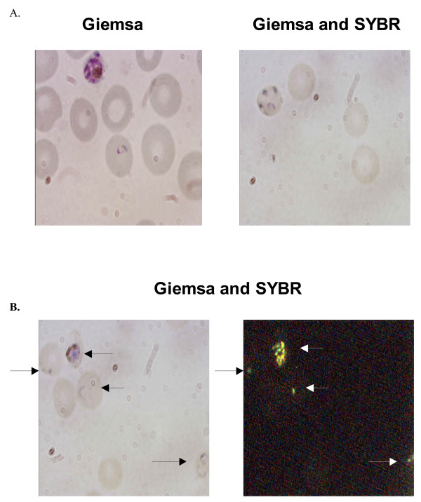
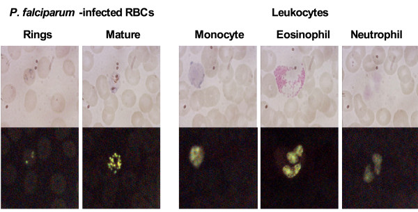
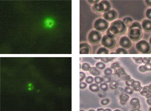
Results: Of the 14 dyes that demonstrated intense fluorescence staining, only SYBR Green 1, YOYO-1 and ethidum homodimer-2 could be detected using fluorescent microscopy, when cells were first stained with Giemsa. Giemsa staining was not effective when applied after the fluorescent dyes. SYBR Green 1 provided the best staining in the presence of Giemsa, as a very high percentage of the parasitized cells were simultaneously stained. When blood films were screened using fluorescence microscopy the parasites were more readily detectable due to the sharp contrast between the dark background and the specific, bright fluorescence produced by the parasites.
Conclusion: The dual staining method reported here allows fluorescence staining, which enhances the reader's ability to detect parasites under low parasitaemia conditions, coupled with the ability to examine the same cell under bright field conditions to detect the characteristic morphology of Plasmodium species that is observed with Giemsa staining.
Figures
Similar articles
-
Flow cytometric enumeration of parasitemia in cultures of Plasmodium falciparum stained with SYBR Green I and CD235A.ScientificWorldJournal. 2014;2014:536723. doi: 10.1155/2014/536723. Epub 2014 Dec 8. ScientificWorldJournal. 2014. PMID: 25548783 Free PMC article.
-
Hydrophilic-treated plastic plates for wide-range analysis of Giemsa-stained red blood cells and automated Plasmodium infection rate counting.Malar J. 2017 Aug 8;16(1):321. doi: 10.1186/s12936-017-1975-9. Malar J. 2017. PMID: 28789644 Free PMC article.
-
Detection of Plasmodium falciparum infection with the fluorescent dye, benzothiocarboxypurine.Am J Trop Med Hyg. 1991 Jan;44(1):11-6. doi: 10.4269/ajtmh.1991.44.11. Am J Trop Med Hyg. 1991. PMID: 1996734
-
Progress and challenges in the use of fluorescence-based flow cytometric assays for anti-malarial drug susceptibility tests.Malar J. 2021 Jan 21;20(1):57. doi: 10.1186/s12936-021-03591-8. Malar J. 2021. PMID: 33478496 Free PMC article. Review.
-
Comparison of acridine orange and giemsa stains for malaria diagnosis.Korean J Parasitol. 1995 Dec;33(4):391-4. doi: 10.3347/kjp.1995.33.4.391. Korean J Parasitol. 1995. PMID: 8591019 Review.
Cited by
-
Malaria theranostics using hemozoin-generated vapor nanobubbles.Theranostics. 2014 May 22;4(7):761-9. doi: 10.7150/thno.9128. eCollection 2014. Theranostics. 2014. PMID: 24883125 Free PMC article.
-
Fluorescence microscope (Cyscope) for malaria diagnosis in pregnant women in Medani Hospital, Sudan.Diagn Pathol. 2011 Sep 24;6:88. doi: 10.1186/1746-1596-6-88. Diagn Pathol. 2011. PMID: 21943212 Free PMC article.
-
Preliminary Evidence of Human Plasmodium in Domestic Animals from a Malaria-Endemic Region in Indonesia.F1000Res. 2024 Dec 10;10:645. doi: 10.12688/f1000research.53946.4. eCollection 2021. F1000Res. 2024. PMID: 39931544 Free PMC article.
-
Rapid and highly sensitive detection of malaria-infected erythrocytes using a cell microarray chip.PLoS One. 2010 Oct 13;5(10):e13179. doi: 10.1371/journal.pone.0013179. PLoS One. 2010. PMID: 20967248 Free PMC article.
-
Image analysis and machine learning for detecting malaria.Transl Res. 2018 Apr;194:36-55. doi: 10.1016/j.trsl.2017.12.004. Epub 2018 Jan 12. Transl Res. 2018. PMID: 29360430 Free PMC article. Review.
References
-
- Kain KC, Harrington MA, Tennyson S, Keystone JS. Imported malaria: prospective analysis of problems in diagnosis and management. Clin Infect Dis. 1998;27:142–149. - PubMed
-
- Christen D, Steffen R, Schlagenhauf P. Deaths caused by malaria in Switzerland 1988-2002. Am J Trop Med Hyg. 2006;75:1188–1194. - PubMed
-
- Jennings RM, JB DES, Todd JE, Armstrong M, Flanagan KL, Riley EM, Doherty JF. Imported Plasmodium falciparum malaria: are patients originating from disease-endemic areas less likely to develop severe disease? A prospective, observational study. Am J Trop Med Hyg. 2006;75:1195–1199. - PubMed
-
- Time for the World Bank to act on malaria. Lancet. 2006;367:1372. - PubMed
Publication types
MeSH terms
Substances
LinkOut - more resources
Full Text Sources
Other Literature Sources
