. 2007;9(4):R67.
doi: 10.1186/ar2226.

Mesangial cells of lupus-prone mice are sensitive to chemokine production

Affiliations

Mesangial cells of lupus-prone mice are sensitive to chemokine production

Shuk-Man Ka et al. Arthritis Res Ther. 2007.

Abstract

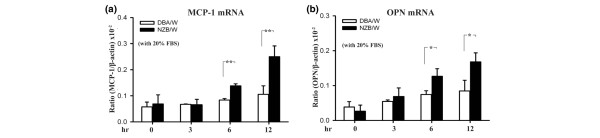
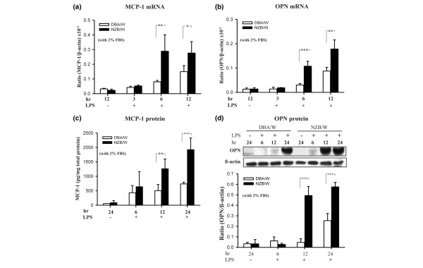
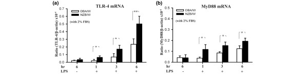
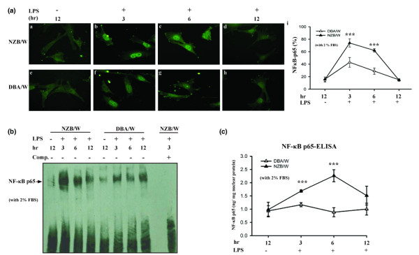
Infectious antigens may be triggers for the exacerbation of systemic lupus erythematosus. The underlying mechanism causing acceleration and exacerbation of lupus nephritis (LN) is largely unknown. Bacterial lipopolysaccharide (LPS) is capable of inducing an accelerated model of LN in NZB/W mice, featuring diffuse proliferation of glomerular resident cells. We hypothesized that mesangial cells (MCs) from LN subjects are more responsive to LPS than normal subjects. Cultured primary NZB/W and DBA/W (nonautoimmune disease-prone strain with MHC class II molecules identical to those of NZB/W) MCs were used. Monocyte chemoattractant protein-1 (MCP-1) and osteopontin (OPN) expressions either in the baseline (normal culture) condition or in the presence of LPS were evaluated by real-time PCR, ELISA, or western blot analysis. NF-kappaB was detected by ELISA, electrophoresis mobility-shift assay, and immunofluorescence. First, either in the baseline condition or in the presence of LPS, NZB/W MCs produced significantly higher levels of MCP-1 and OPN than the DBA/W MC controls. Second, NZB/W MCs expressed significantly higher levels of Toll-like receptor 4, myeloid differentiation factor 88, and NF-kappaB than the DBA/W MC controls, both receiving exactly the same LPS treatment. In conclusion, NZB/W MCs are significantly more sensitive than their normal control DBA/W MCs in producing both MCP-1 and OPN. With LPS treatment, the significantly elevated levels of both chemokines produced by NZB/W MCs are more likely due to a significantly greater activation of the Toll-like receptor 4-myeloid differentiation factor 88-associated NF-kappaB pathway. The observed abnormal molecular events provide an intrarenal pathogenic pathway involved in an accelerated type of LN, which is potentially infection triggered.


Figures

Figure 1
Figure 1
Monocyte chemoattractant protein-1 and osteopontin mRNA expression in NZB/W mesangial cells under the baseline condition. Mesangial cells were incubated in the baseline (normal culture, 20% fetal bovine serum (FBS)) condition for different periods of time, and then levels of (a) monocyte chemoattractant protein-1 (MCP-1) mRNA or (b) osteopontin (OPN) mRNA were examined by real-time PCR. The experiment was performed in triplicate, and results are expressed as the mean ± standard error, n = 6. *P < 0.05, **P < 0.01.
Figure 2
Figure 2
Monocyte chemoattractant protein-1 and osteopontin expression in NZB/W mesangial cells with lipopolysaccharide treatment. Growth-arrested (under 2% fetal bovine serum (FBS)) mesangial cells were incubated with 10 μg/ml lipopolysaccharide (LPS) for different periods of time, and then levels of (a) monocyte chemoattractant protein-1 (MCP-1) mRNA or (b) osteopontin (OPN) mRNA were examined by real-time PCR. (c) MCP-1 protein levels in the culture supernatant were measured by ELISA. (d) OPN protein levels in the cell lysate were measured by western blot analysis; semiquantitative data are shown. The MCP-1 protein expression levels at various time points were normalized to total protein (pg/mg). The experiment was performed in triplicate, and results are expressed as the mean ± standard error, n = 6. The last time point without LPS stimulation (unstimulated) was presented as the negative control. *P < 0.05, **P < 0.01, ***P < 0.005.
Figure 3
Figure 3
Toll-like receptor 4 and myeloid differentiation factor 88 mRNA in NZB/W mesangial cells (lipopolysaccharide treatment). Growth-arrested (under 2% fetal bovine serum (FBS)) mesangial cells were incubated with 10 μg/ml lipopolysaccharide (LPS) for different periods of time, and then Toll-like receptor 4 (TLR-4) and myeloid differentiation factor 88 (MyD88) mRNA levels were measured by real-time PCR analysis. The experiment was performed in triplicate, and results are expressed as the mean ± standard error, n = 6. The last time point without LPS stimulation (unstimulated) was presented as the negative control. *P < 0.05, **P < 0.01.
Figure 4
Figure 4
NF-κB p65 activation in NZB/W mesangial cells with lipopolysaccharide treatment. Growth-arrested (under 2% fetal bovine serum (FBS)) mesangial cells (MCs) were incubated with 10 μg/ml lipopolysaccharide (LPS) for different periods of time, and then the distribution of NF-κB p65 was examined. (a) Immunofluorescence: images a–d, NZB/W MCs; images e–h, DBA/W MCs (nonimmune strain, served as control) incubated for 0–12 hours with LPS; and image i, semiquantitative data. (b) Electrophoresis mobility-shift assay performed using a DIG-labeled synthetic oligonucleotide and nuclear extract from MCs. The competition assay used the same unlabeled oligonucleotide at a 10-fold higher concentration. Arrow, NF-κB p65 binding bands. Comp., the abbreviation of competition. (c) ELISA performed using the TransAM NF-κB p65 kit. The NF-κB p65 expression levels at various time points were normalized to nuclear protein (ng/mg). The experiment was performed in triplicate, and results are expressed as the mean ± standard error, n = 6. The last time point without LPS stimulation (unstimulated) was presented as the negative control. ***P < 0.005 versus DBA/W MC controls.
Figure 5
Figure 5
NF-κB inhibitor effect on lipopolysaccharide-induced monocyte chemoattractant protein-1 and osteopontin expression in NZB/W mesangial cells. Growth-arrested (under 2% fetal bovine serum (FBS)) mesangial cells were preincubated for 2 hours with n-tosyl-1-phenylalanine chloromethyl ketone (TPCK) or dexamethasone (Dex), and then 10 μg/ml lipopolysaccharide (LPS) was added for 12 hours: (a) monocyte chemoattractant protein-1 (MCP-1) mRNA levels and (b) osteopontin (OPN) mRNA levels were measured by real-time PCR analysis. Growth-arrested (under 2% FBS) mesangial cells were preincubated for 2 hours with TPCK or Dex, then 10 μg/ml LPS was added for 24 hours: (c) MCP-1 protein levels in the supernatant were measured by ELISA, and (d) OPN protein levels in the cell lysate were measured by western blot analysis; semiquantitative data are shown. The MCP-1 protein expression levels at various time points were normalized to total protein (pg/mg). The experiment was performed in triplicate, and results are expressed as the mean ± standard error. **P < 0.01, ***P < 0.005, n = 6. White bar, absence of LPS; black bar, LPS alone; hatched bar, TPCK and LPS or Dex and LPS.

Similar articles

Cited by

References

    1. Waldman M, Madaio MP. Pathogenic autoantibodies in lupus nephritis. Lupus. 2005;14:19–24. doi: 10.1191/0961203305lu2054oa. - DOI - PubMed
    1. Zandman-Goddard G, Shoenfeld Y. Infections and SLE. Autoimmunity. 2005;38:473–485. doi: 10.1080/08916930500285352. - DOI - PubMed
    1. Cavallo T, Granholm NA. Accelerated (proliferative) lupus nephritis. Am J Pathol. 1990;137:1549–1551. - PMC - PubMed
    1. Cavallo T, Granholm NA. Bacterial lipopolysaccharide transforms mesangial into proliferative lupus nephritis without interfering with processing of pathogenic immune complexes in NZB/W mice. Am J Pathol. 1990;137:971–978. - PMC - PubMed
    1. Wang Y, Rangan GK, Goodwin B, Tay YC, Harris DC. Lipopolysaccharide-induced MCP-1 gene expression in rat tubular epithelial cells is nuclear factor-kappaB dependent. Kidney Int. 2000;57:2011–2022. doi: 10.1046/j.1523-1755.2000.00051.x. - DOI - PubMed

Publication types

MeSH terms