. 2007 Aug 31;360(3):659-65.
doi: 10.1016/j.bbrc.2007.06.106. Epub 2007 Jun 28.

PLAGL2 translocation and SP-C promoter activity--a cellular response of lung cells to hypoxia

Affiliations

PLAGL2 translocation and SP-C promoter activity--a cellular response of lung cells to hypoxia

Yuhong Guo et al. Biochem Biophys Res Commun. .

Abstract

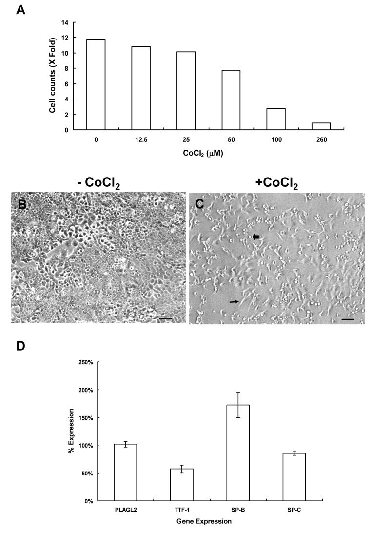
Cobalt is a transition metal which can substitute for iron in the oxygen-sensitive protein and mimic hypoxia. Cobalt was known to be associated with the development of lung disease. In this study, when lung cells were exposed to hypoxia-induced by CoCl(2) at a sub-lethal concentration (100 microM), their thyroid transcription factor-1 (TTF-1) expression was greatly reduced. Under this condition, SP-B promoter activity was down-regulated, but SP-C promoter remained active. Therefore, we hypothesized that other factor(s) besides TTF-1 might contribute to the modulation of SP-C promoter in hypoxic lung cells. Pleomorphic adenoma gene like-2 (PLAGL2), a previously identified TTF-1-independent activator of the SP-C promoter, was not down-regulated, nor increased, within those cells. Its cellular location was redistributed from the cytoplasm to the nucleus. Chromatin immunoprecipitation (ChIP) and quantitative RT-PCR analyses demonstrated that nuclear PLAGL2 occupied and transactivated the endogenous SP-C promoter in lung cells. Thereby, through relocating and accumulating of PLAGL2 inside the nucleus, PLAGL2 interacted with its target genes for various cellular functions. These results further suggest that PLAGL2 is an oxidative stress responding regulator in lung cells.


Figures

Figure 1
Figure 1
MLE12 cells growth rate and TTF-1 expression were suppressed by CoCl2-induced hypoxia. (A) The cell number changes of MLE12 cells were graphed against various concentrations of CoCl2 in medium. Cell counts in duplicated wells from two individual experiments were averaged and plotted. Fold of cell number increase was compared to the cell number in the overnight culture before treated with CoCl2. (B and C) The growth and cell morphology of MLE12 cells were changed by CoCl2 treatment. Equal amounts of cells (2X10⁴/well) were seeded overnight before treated with CoCl2. After 48 hours of treatment, cells without (B) or with (C) the CoCl2 treatment were observed under microscope with phase contrast. Arrow and blockarrow in C denoted cells with morphological changes to spindle or spherical and lose adhering. The size bar indicated 100 μm. Magnification 10X. (D) Gene expression pattern in MLE12 cells treated with 100 μM CoCl2 for 48 hours. Total RNA from harvested cells was isolated and analyzed for the expression of indicated genes. Quantitative RT-PCR analysis was performed to measure and compare the level of individual gene transcript in CoCl2 treated and control samples. The expression level in the control was set at 100%. Data collected for statistical analysis was as described (N≥12).
Figure 2
Figure 2
SP-C promoter activity remained in CoCl2 treated H441 cells. (A). PLAGL2 expression was not changed in CoCl treated H441 cells. A total of 4X10⁴ cells/well in 24-well plate were seeded 48 hours before being treated with various amounts of CoCl2 in culture medium. Cells were harvested after 48 hours treatment, lysed, western blotted, and probed with mAb85C47-1, anti-TTF-1, and anti-actin antibodies sequentially. (B). The SP-C promoter remained active in 100 μM CoCl2 treated H441 cells. Cells as described in panel A were transfected with pGL3-hSP-BP (closed bar, 50 ng/well) or pGL3-hSP-CP (open bar, 400 ng/well) promoter with various concentrations of CoCl2 as labeled. Luciferase activities in cells were measured after 48 hours of transfection and statistically analyzed. Reporter gene activities in control without CoCl2 treatment were set to 100%. The difference of luciferase activities between SP-B and SP-C promoters in 100 μM CoCl2 treated cells was significant (**: p<0.01, N=12)
Figure 3
Figure 3
CoCl2 induced PLAGL2 translocation into the nuclei of H441 cells. H441 cells without (A and B) or with (C and D) CoCl2 (100 μM) treatment were fixed and probed with mAb85C47-1 (B and D). PLAGL2 was distributed in the cytoplasm and the nucleus compartments (B, arrows) or around the perinuclear region (B, blockarrow) of H441 cells. After adding CoCl2 for 48 hours, PLAGL2 signal was observed more intense in the nuclei (D, arrows). The size bar represented 100 μm. Magnification (20X).
Figure 4
Figure 4
PLAGL2 is associated with the SP-C promoter in H441 and transfected MLE12 cells. H441 (A) or Flag-PLAGL2 transfected MLE12 cells (B) were fixed in situ for the ChIP analysis. The harvested chromatin were immunoprecipitated with various antibodies as indicated above the panel. PCR was used to detect the SP-C and SP-B promoters within the precipitated chromatin associated with those antigen-antibody complexes. PCR amplified DNA fragments were visualized in a 2% agarose gel. The arrow points to the amplified SP-C or SP-B promoter DNA fragment in (B). The association of PLAGL2 to the SP-C promoter increases endogenous SP-C expression in transfected MLE12 cells (C). MLE12 cells transfected with 400 ng of pCIN-Flag (open bar) or Flag-PLAGL2 (closed bar) were incubated for 48 hours, harvested, and then subjected for gene expression evaluation by quantitative RT-PCR analysis. A t-test (N=5) was performed by comparing the expression of SP-B or SP-C genes in PLAGL2 transfected cells and pCIN-Flag vector transfected cells. The value for the control cells was set to 100%. (**: p<0.01).

Similar articles

Cited by

References

    1. Kelly SE, Bachurski CJ, Burhans MS, Glasser SW. Transcription of the lung-specific surfactant protein C gene is mediated by thyroid transcription factor 1. J Biol.Chem. 1996;271:6881–6888. - PubMed
    1. Bohinski RJ, Di Lauro R, Whitsett JA. The lung-specific surfactant protein B gene promoter is a target for thyroid transcription factor 1 and hepatocyte nuclear factor 3, indicating common factors for organ-specific gene expression along the foregut axis. Mol.Cell Biol. 1994;14:5671–5681. - PMC - PubMed
    1. Yang MC, Weissler JC, Terada LS, Deng F, Yang YS. Pleiomorphic Adenoma Gene-Like-2, a Zinc Finger Protein, Transactivates the Surfactant Protein-C Promoter. Am.J.Respir.Cell Mol.Biol. 2005;32:35–43. - PubMed
    1. Kas K, Voz ML, Hensen K, Meyen E, Van de Ven WJ. Transcriptional activation capacity of the novel PLAG family of zinc finger proteins. J.Biol Chem. 1998;273:23026–23032. - PubMed
    1. Hensen K, Van Valckenborgh IC, Kas K, Van de Ven WJ, Voz ML. The tumorigenic diversity of the three PLAG family members is associated with different DNA binding capacities. Cancer Res. 2002;62:1510–1517. - PubMed

Publication types

MeSH terms

LinkOut - more resources