Review
. 2007 Oct-Nov;42(4-5):363-71.
doi: 10.1016/j.ceca.2007.05.007. Epub 2007 Jul 5.

Homer proteins in Ca2+ signaling by excitable and non-excitable cells

Affiliations
Review

Homer proteins in Ca2+ signaling by excitable and non-excitable cells

Paul F Worley et al. Cell Calcium. 2007 Oct-Nov.

Abstract

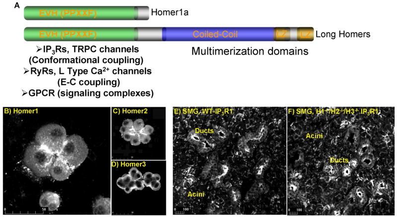
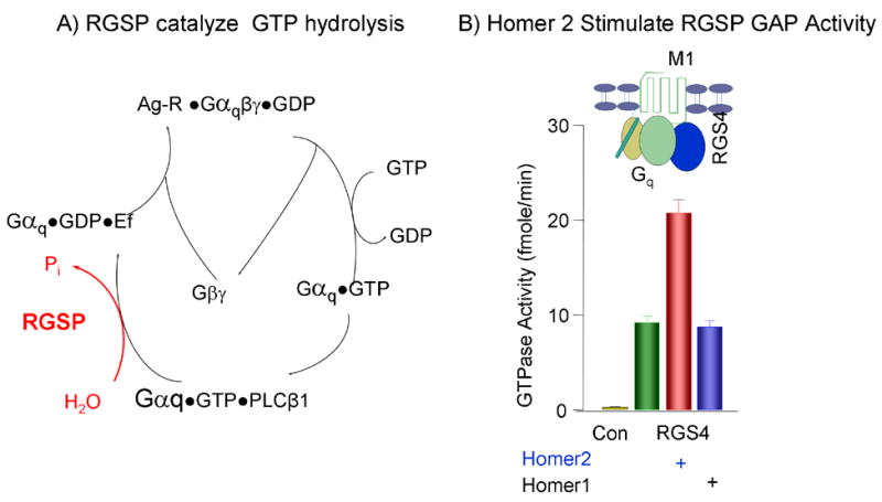
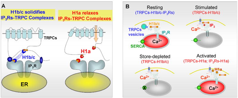
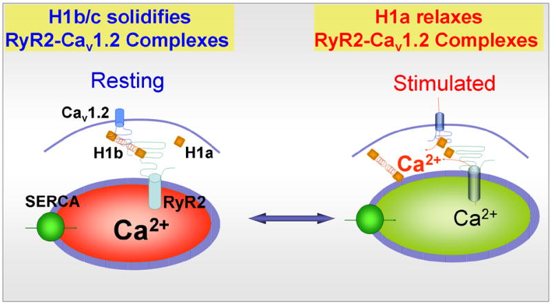
Homers are scaffolding proteins that bind Ca(2+) signaling proteins in cellular microdomains. The Homers participate in targeting and localization of Ca(2+) signaling proteins in signaling complexes. However, recent work showed that the Homers are not passive scaffolding proteins, but rather they regulate the activity of several proteins within the Ca(2+) signaling complex in an isoform-specific manner. Homer2 increases the GAP activity of RGS proteins and PLCbeta that accelerate the GTPase activity of Galpha subunits. Homer1 gates the activity of TRPC channels, controls the rates of their translocation and retrieval from the plasma membrane and mediates the conformational coupling between TRPC channels and IP(3)Rs. Homer1 stimulates the activity of the cardiac and neuronal L-type Ca(2+) channels Ca(v)1.2 and Ca(v)1.3. Homer1 also mediates the communication between the cardiac and smooth muscle ryanodine receptor RyR2 and Ca(v)1.2 to regulate E-C coupling. In many cases the Homers function as a buffer to reduce the intensity of Ca(2+) signaling and create a negative bias that can be reversed by the immediate early gene form of Homer1. Hence, the Homers should be viewed as the buffers of Ca(2+) signaling that ensure a high spatial and temporal fidelity of the Ca(2+) signaling and activation of downstream effects.


Figures

Fig. 1
Fig. 1
Panel (A) shows the Homer domains and known interacting Ca2+ signaling proteins. Isoform-specific localization of Homer1 (B), Homer2 (C) and Homer3 (D) is demonstrated in pancreatic acini. Similar localization of type 1 IP3R in observed in WT cells (E) and cells from which all Homer isoforms were deleted. Panels (B–D) are reproduced from [30] with permission.
Fig. 2
Fig. 2
Panel (A) depicts the turn over cycle of Gq is response to receptor stimulation and the function of RGS proteins (RGSP) as Gαq GTPase activating proteins (GAP). Panel (B) show the stimulation of RGS4 GAP activity by Homer2, but not by Homer1, in reconstituted microsomes composed of Gq, the M1 receptor and RGS4 and stimulated with carbachol. The results in (B) were taken from [30] with permission.
Fig. 3
Fig. 3
Panel (A) shows a model of a potential mechanism by which Homer1 gates TRPC channels. TRPC channels and IP3Rs have Homer binding ligands. In resting state, TRPC channels are bound to the long H1b/c through their single (all TRPCs, except TRPC1) or two (TRPC1) Homer binging ligands and to the IP3Rs. In this complex, H1b/c solidifies interaction between the channels and the channels are not active. In stress, H1a is up-regulated and replaces H1b/c. This results in relaxation of the interaction between the channels to prevent inhibition of TRPC channels activity by the IP3Rs, resulting in spontaneously active TRPC channels. Panel (B) shows a model of a potential mechanism for regulation of TRPC channels translocation by Homer1. The intracellular, vesicular pool of TRPC channels exists in TRPCs-H1b/c-IP3Rs complexes. Cell stimulation generates high level of IP3. Binding of IP3 to the IP3Rs dissociate the binding of IP3Rs to H1b/c, but H1b/c remains bound to the TRPCs. This results in activation of TRPC channels and allows the rapid re-binding of IP3Rs to H1b/c and reassembly of the complex upon termination of cell stimulation. Store depletion and H1a can also dissociate the TRPCs-H1b/c-IP3 receptors complexes to activate TRPC channels and Ca2+ influx.
Fig. 4
Fig. 4
Homer1 regulates E–C coupling in a mechanism equivalent to that of conformational-coupling found for TRPC channels. H1b/c binds Cav1.2 (and Cav1.3 in neurons) and RyR2 to form the complex Cav1.2-H1b/c-RyR2. In this complex H1b/c solidifies the interaction between the channels to hinder the conformational transmission between Cav1.2 and RyR2 in response to membrane depolarization and reduce muscle (and neuronal) excitability. When H1a is up-regulated, it binds to the channels to relax the interaction between Cav1.2 and RyR2 and facilitate the conformational transmission between Cav1.2 and RyR2. Furthermore, H1a activates Cav1.2 (and Cav1.3) to further enhance Ca2+ influx and activation of CICR. The two effects of H1a result in increase muscle (and neuronal) excitability and increasing spontaneous contraction (and perhaps firing of spontaneous action potentials).

References

    1. Kato A, Ozawa F, Saitoh Y, Fukazawa Y, Sugiyama H, Inokuchi K. Novel members of the Vesl/Homer family of PDZ proteins that bind metabotropic glutamate receptors. J Biol Chem. 1998;273:23969–75. - PubMed
    1. Brakeman PR, Lanahan AA, O'Brien R, Roche K, Barnes CA, Huganir RL, Worley PF. Homer: a protein that selectively binds metabotropic glutamate receptors. Nature. 1997;386:284–8. - PubMed
    1. Xiao B, Tu JC, Worley PF. Homer: a link between neural activity and glutamate receptor function. Curr Opin Neurobiol. 2000;10:370–4. - PubMed
    1. Soloviev MM, Ciruela F, Chan WY, McIlhinney RA. Molecular characterisation of two structurally distinct groups of human homers, generated by extensive alternative splicing. J Mol Biol. 2000;295:1185–200. - PubMed
    1. Tadokoro S, Tachibana T, Imanaka T, Nishida W, Sobue K. Involvement of unique leucine-zipper motif of PSD-Zip45 (Homer 1c/vesl-1L) in group 1 metabotropic glutamate receptor clustering. Proc Natl Acad Sci U S A. 1999;96:13801–6. - PMC - PubMed