. 2007 Oct 25;367(2):398-412.
doi: 10.1016/j.virol.2007.05.002. Epub 2007 Jul 9.

The vaccinia virus E8R gene product is required for formation of transcriptionally active virions

Affiliations

The vaccinia virus E8R gene product is required for formation of transcriptionally active virions

Sayuri E M Kato et al. Virology. .

Abstract

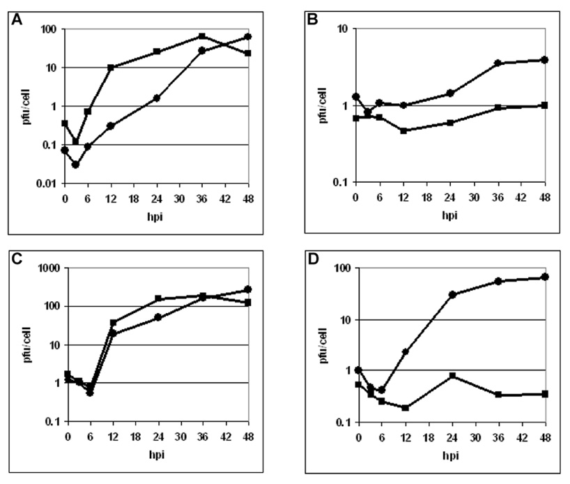
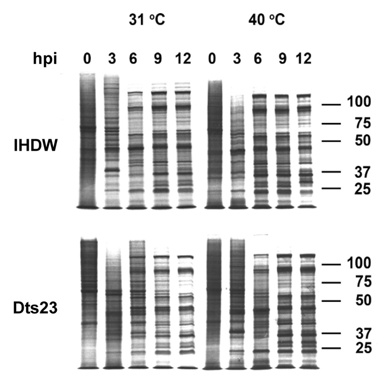
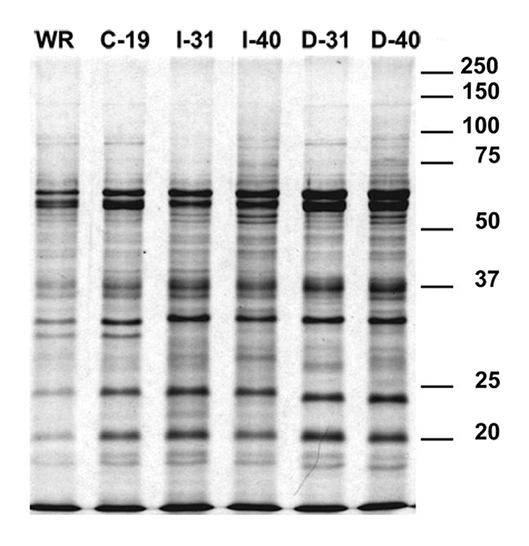
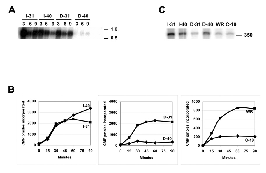
Two vaccinia virus temperature-sensitive mutants were mapped to the E8R gene and subjected to phenotypic characterization. Dts23 contains a missense mutation in the coding region of E8R (L81F), and in Cts19 the initiating methionine codon is changed from ATG to ATA (M1I). The two ts mutants display normal patterns of gene expression and DNA replication during infection. The E8 protein is synthesized exclusively late during infection and packaged into virion cores Western blot analysis revealed that E8 synthesis is reduced in Dts23 infected cells at permissive (31 degrees C) and non-permissive temperature (39.7 degrees C) and absent in Cts19 infection under both conditions. Dts23 virions produced at 39.7 degrees C were indistinguishable in appearance from wt virions. Cts19 fails to produce identifiable viral structures when incubated at 39.7 degrees C. Purified Dts23 virions produced at 39.7 degrees C contain reduced amounts of E8 and have a high particle to infectivity ratio; purified Cts19 virions grown at 31 degrees C also show reduced infectivity and do not contain detectable E8. Dts23 grown at 39.7 degrees C could enter cells but failed to synthesize early mRNA or produce CPE. Soluble extracts from mutant virions were active in a promoter dependent in vitro transcription assay, however intact mutant cores were defective in transcription. We suggest that E8 plays a subtle role in virion core structure that impacts directly or indirectly on core transcription.


Figures

Fig. 1
Fig. 1. One-step marker rescue of E8R mutants
BSC40 cells were infected with Dts23 or Cts19 mutant virus and transfected with a PCR product as described in Methods. Crystal violet-stained dishes are shown, labeled with the PCR amplification used in the transfection as shown in the drawing on the top of the Figure. (A): Dts23; (B): Cts19.
Fig. 2
Fig. 2. One-step growth of wt, Dts23 and Cts19 viruses
BSC40 cells were infected at moi = 10 pfu/cell with wt or mutant viruses, incubated at 31 or 39.7 °C for the indicated periods of time (abscissa), and virus yields were determined by plaque titration at 31°C (ordinate). (A) WR-wt; (B) Cts19; (C) IHDW-wt; (D) Dts23. (31°C - ●; 39.7°C - ■)
Fig. 3
Fig. 3. Viral DNA replication in virus infected cells
BSC-40 cells were infected at moi=10 pfu/cell, incubated at 31 or 39.7 °C, and at indicated times postinfection, cell extracts were prepared and applied on nylon membranes, and hybridization to a 32P labeled vaccinia virus DNA probe was performed as described under Methods. The numbers (arbitrary units) express the average value of three different measurements. (A) IHDW-wt; (B) Dts23. (31°C - ●; 39.7°C - ■)
Fig. 4
Fig. 4. Protein synthesis in wt and mutant-infected cells
BSC40 cells were infected at moi = 10 pfu/cell, incubated at 31 or 39.7 °C, and pulse-labeled with 35S methionine at various times postinfection, indicated in hours at the top of each autoradiogram. Cells lysates were electrophoresed on SDS-PAGE, and gels were dried and autoradiographed. Incubation temperature and time of the pulse label is indicated at the top of each column, and the mutant used in the infection is indicated to the left of each row of autoradiograms. Approximate molecular weights, in kDa, are indicated to the right of each autoradiograms.
Fig. 5
Fig. 5. Electron micrographs of wt and mutant-infected cells
BSC40 cells were infected with wt or mutant virus at a moi = 10 pfu/cell, incubated at 31 or 39.7 °C for 16 hs (D); 24 hs (A–C, E, G; inset); 36 hs (H) and 48 hs (F) and processed for electron microscopy as described in Methods. IV= immature virions; MV= mature virions; WV= wrapped virions; EV= extracellular virions; C= crescents; N= nucleus; F= DNA factories. (A) Dts23 infections at 31 °C; (B – D) Dts23 infections at 39.7 °C; (E – F) Cts19 infections at 31 °C; (G – H) Cts19 infections at 39.7 °C. The inset in C shows the formation of WV and EV in Dts23 infections. Bar= 1 µm
Fig. 6
Fig. 6. Accumulation of E8 in wt and mutant virus infected cells
BSC40 cells were infected with wt or mutant virus at a moi = 10 pfu/cell and incubated at 31 or 39.7 °C. At different times, indicated in hours on the top of each lane, samples were removed and processed for Western blot as described under Methods. The arrow represents the expected MW for the E8 protein. Approximate molecular weights, in kDa, are indicated to the right of the F.
Fig. 7
Fig. 7. Kinetics of synthesis of E8
BSC40 cells were infected with VACV-WR at a moi = 10 pfu/cell and incubated at 37 °C in the absence (C) or in the presence of 40 µg/ml of CAR (A). At 1, 2, 3, 5, 6, and 8 hours post-infection samples were removed and prepared for Western blot as described in Methods. M and MA are mock infected cells incubated in the absence or presence of CAR. Approximate molecular weights, in kDa, are indicated to the right of the autoradiograms.
Fig. 8
Fig. 8. Analysis of proteins of purified wt and mutants virions
0.18 A260nm units (12 µg) of purified virions were resuspend in SDS-sample buffer, the proteins were separated in an 11% SDS-PAGE and stained with Coomassie blue as described in Methods. The virion analyzed in labeled on the top of each lane: WR, wt; C-19, Cts19 grown at 31°C; I-31, IHDW grown at 31 °C; I-40, IHDW grown at 39.7 °C; D-31, Dts23 grown at 31 °C; D-40, Dts23 grown at 39.7 °C. Molecular weights markers are indicated to the right of the gel, in kDa.
Fig. 9
Fig. 9. Western blot analysis of virions core and membrane fractions
0.06 A260nm units (0.7 µg) of each virion sample, labeled as described in Fig.8, was resuspended in core buffer and processed as described under Methods. The gene product corresponding to each antiserum used is indicated to the left of each row; the virus sample probed is indicated on the top of each lane where P= pellet/core fraction and S= soluble/membrane fraction; the molecular weight markers are indicated on the right side of the blot, in kDa.
Fig. 10
Fig. 10. Analysis of entry process of wt and mutants
BSC40 cells were infected with moi = 1000 particles of each virus grown at 31 °C (A, C) or 39.7 °C (B, D, F), fixed, permeabilized, stained for the viral core protein A4 (green) and for DNA(blue), and analyzed by confocal microscopy as described in Methods. IHDW= A, B; Dts23= C, D, F; mock infected (E).
Fig. 11
Fig. 11. In vivo and in vitro RNA synthesis of purified virions
A) Northern blot analysis of wt and mutants infected cells. BSC40 cells were infected with the indicated virus at moi = 1000 particles per cell and incubated at 31 °C. At 3, 6 and 9 post-infection samples were removed and prepared for Northern blot. Following pre-hybridization, 1 × 10⁷ cpm of a riboprobe, synthesized from the C11R gene, was added to the blot and incubated overnight at 55 °C as described in Methods. The virion analyzed in labeled on the top of each lane: WR, wt; C-19, Cts19 grown at 31°C; I-31, IHDW grown at 31 °C; I-40, IHDW grown at 39.7 °C; D-31, Dts23 grown at 31 °C; D-40, Dts23 grown at 39.7 °C; the molecular weight markers in the right in kb. B) RNA synthesis directed by purified wt and mutants particles. Purified virions were permeabilized with NP40 and DTT treatment, incubated with [α32P] CTP and the other nucleotides for 90 minutes. At indicated times, samples were removed, and the acid precipitable radioactivity was determined as described in Methods. The virions were labeled as above. C) Specific early transcription directed by wt and mutant virion extracts. Soluble transcription extracts were prepared by incubating purified VACV cores with sodium deoxycholate and passing through a DEAE-cellulose column. The active fractions were pooled, assayed for specific early transcription, and the product analyzed by electrophoresis on denaturing polyacrylamide gel as described in Methods. The virions were labeled as above; the molecular weight marker in indicated at the right in nucleotides.

Similar articles

Cited by

References

    1. Ausubel FM, Brent R, Kingston RE, Moore DD, Seidman JG, Smith JA, Struhl K. Current protocols in molecular biology. New York: John Wiley & Sons; 1994.
    1. Bayliss CD, Smith GL. Vaccinia virion protein VP8, the 25 kDa product of the L4R gene, binds single-stranded DNA and RNA with similar affinity. Nucleic Acids Res. 1997;25:3984–3990. - PMC - PubMed
    1. Berger SL. Histone modifications in transcriptional regulation. Curr. Opin. Genet. Dev. 2002;12:142–148. - PubMed
    1. Black EP, Moussatche N, Condit RC. Characterization of the interactions among vaccinia virus transcription factors G2R, A18R, and H5R. Virology. 1998;245:313–322. - PubMed
    1. Broyles SS. Vaccinia virus transcription. J. Gen. Virol. 2003;84:2293–2303. - PubMed

Publication types