

Immunohistological determination of ecto-nucleoside triphosphate diphosphohydrolase1 (NTPDase1) and 5'-nucleotidase in rat hippocampus reveals overlapping distribution
- PMID: 17619139
- PMCID: PMC11517217
- DOI: 10.1007/s10571-007-9159-8
Immunohistological determination of ecto-nucleoside triphosphate diphosphohydrolase1 (NTPDase1) and 5'-nucleotidase in rat hippocampus reveals overlapping distribution
Abstract
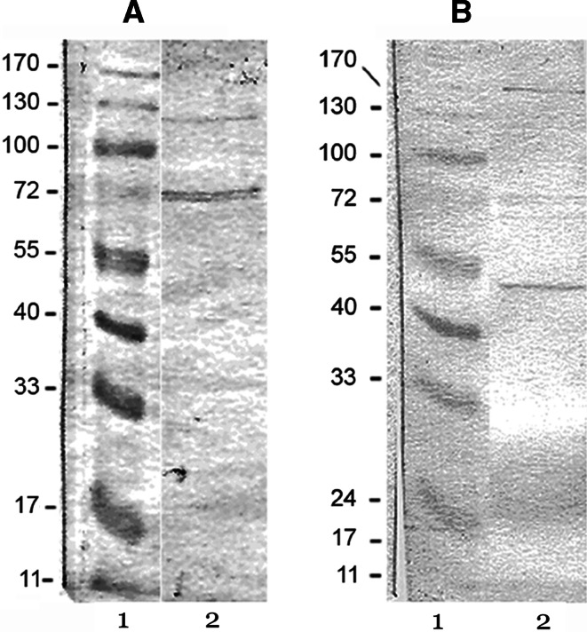
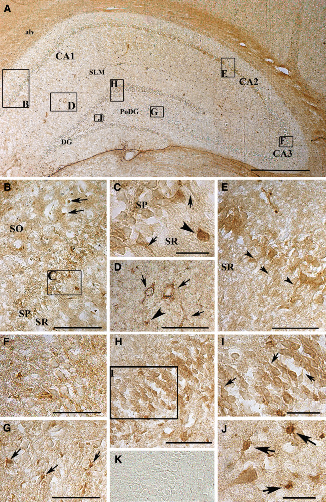
Distribution of two enzymes involved in the ectonucleotidase enzyme chain, ecto-nucleoside triphosphate diphosphohydrolase1 (NTPDase1) and ecto-5'-nucleotidase, was assessed by immunohistochemistry in the rat hippocampus. Obtained results have shown co-expression of the enzymes in the hippocampal region, as well as wide and strikingly similar cellular distribution. Both enzymes were expressed at the surface of pyramidal neurons in the CA1 and CA2 sections, while cells in the CA3 section were faintly stained. The granule cell layer of the dentate gyrus was moderately stained for NTPDase1, as well as for ecto-5'-nucleotidase. Glial association for ecto-5'-nucleotidase was also observed, and fiber tracts were intensively stained for both enzymes. This is the first comparative study of NTPDase1 and ecto-5'-nucleotidase distribution in the rat hippocampus. Obtained results suggest that the broad overlapping distribution of these enzymes in neurons and glial cells reflects the functional importance of ectonucleotidase actions in the nervous system.
Figures

Similar articles
-
Spatial Distribution and Expression of Ectonucleotidases in Rat Hippocampus After Removal of Ovaries and Estradiol Replacement.Mol Neurobiol. 2019 Mar;56(3):1933-1945. doi: 10.1007/s12035-018-1217-3. Epub 2018 Jul 6. Mol Neurobiol. 2019. PMID: 29978426
-
Upregulation of nucleoside triphosphate diphosphohydrolase-1 and ecto-5'-nucleotidase in rat hippocampus after repeated low-dose dexamethasone administration.J Mol Neurosci. 2015 Apr;55(4):959-67. doi: 10.1007/s12031-014-0452-y. Epub 2014 Nov 1. J Mol Neurosci. 2015. PMID: 25367797
-
Immunolocalization of ectonucleotidases along the rat nephron.Am J Physiol Renal Physiol. 2006 Feb;290(2):F550-60. doi: 10.1152/ajprenal.00151.2005. Epub 2005 Sep 27. Am J Physiol Renal Physiol. 2006. PMID: 16189292
-
Therapeutic potentials of ecto-nucleoside triphosphate diphosphohydrolase, ecto-nucleotide pyrophosphatase/phosphodiesterase, ecto-5'-nucleotidase, and alkaline phosphatase inhibitors.Med Res Rev. 2014 Jul;34(4):703-43. doi: 10.1002/med.21302. Epub 2013 Sep 23. Med Res Rev. 2014. PMID: 24115166 Review.
-
Ecto-5'-nucleotidase in B-cell chronic lymphocytic leukemia.Biomed Pharmacother. 2002 Mar;56(2):100-4. doi: 10.1016/s0753-3322(01)00072-5. Biomed Pharmacother. 2002. PMID: 12000134 Review.
Cited by
-
Role of Ectonucleotidases in Synapse Formation During Brain Development: Physiological and Pathological Implications.Curr Neuropharmacol. 2019;17(1):84-98. doi: 10.2174/1570159X15666170518151541. Curr Neuropharmacol. 2019. PMID: 28521702 Free PMC article. Review.
-
Developmental increase in ecto-5'-nucleotidase activity overlaps with appearance of two immunologically distinct enzyme isoforms in rat hippocampal synaptic plasma membranes.J Mol Neurosci. 2014 Sep;54(1):109-18. doi: 10.1007/s12031-014-0256-0. Epub 2014 Feb 22. J Mol Neurosci. 2014. PMID: 24563227
-
Enzyme histochemistry: a useful tool for examining the spatial distribution of brain ectonucleotidases in (patho)physiological conditions.Histol Histopathol. 2022 Oct;37(10):919-936. doi: 10.14670/HH-18-471. Epub 2022 May 16. Histol Histopathol. 2022. PMID: 35575291 Review.
-
ATP hydrolysis pathways and their contributions to pial arteriolar dilation in rats.Am J Physiol Heart Circ Physiol. 2011 Oct;301(4):H1369-77. doi: 10.1152/ajpheart.00556.2011. Epub 2011 Jul 29. Am J Physiol Heart Circ Physiol. 2011. PMID: 21803949 Free PMC article.
-
Expression of ecto-nucleoside triphosphate diphosphohydrolase1-3 (NTPDase1-3) by cortical astrocytes after exposure to pro-inflammatory factors in vitro.J Mol Neurosci. 2013 Nov;51(3):871-9. doi: 10.1007/s12031-013-0088-3. Epub 2013 Aug 29. J Mol Neurosci. 2013. PMID: 23990338
References
-
- Almeida T, Rodrigues RJ, de Mendonca A, Ribeiro JA, Cunha RA (2003) Purinergic P2 receptors trigger adenosine release leading to adenosine A2A receptor activation and facilitation of long-term potentiation in rat hippocampal slices. Neuroscience 122:111–121 - PubMed
-
- Belcher SM, Zsarnovszsky A, Crawford PA, Hemani H, Spurling L, Kirley TL (2006) Immunolocalization of ecto-nucleoside triphosphate diphosphohydrolase 3 in rat brain: implications for modulation of multiple homeostatic systems including feeding and sleep-wake behaviors. Neuroscience 137:1331–1346 - PubMed
-
- Bernstein HG, Weiss J, Luppa H (1978) Cytochemical investigations on the localization of 5′-nucleotidase in the rat hippocampus with special reference to synaptic regions. Histochemistry 55:261–267 - PubMed
-
- Bjelobaba I, Nedeljkovic N, Subasic S, Lavrnja I, Pekovic S, Stojkov D, Rakic L, Stojiljkovic M (2006) Immunolocalization of ecto-nucleotide pyrophosphatase/phosphodiesterase 1 (NPP1) in the rat forebrain. Brain Res 1120:53–64 - PubMed
-
- Boeck CR, Sarkis JJ, Vendite D (2002) Kinetic characterization and immunodetection of ecto-ATP diphosphohydrolase (EC 3.6.1.5) in cultured hippocampal neurons. Neurochem Int 40:449–453 - PubMed
Publication types
MeSH terms
Substances
LinkOut - more resources
Full Text Sources
Research Materials
Miscellaneous
