

What is new in pediatric cardiac imaging?
- PMID: 17619900
- PMCID: PMC2668620
- DOI: 10.1007/s00431-007-0544-6
What is new in pediatric cardiac imaging?
Abstract
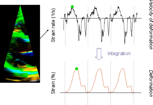
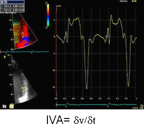
Cardiac imaging has had significant influence on the science and practice of pediatric cardiology. Especially the development and improvements made in non-invasive imaging techniques, like echocardiography and cardiac magnetic resonance imaging (MRI), have been extremely important. Technical advancements in the field of medical imaging are quickly being made. This review will focus on some of the important evolutions in pediatric cardiac imaging. Techniques such as intracardiac echocardiography, 3D echocardiography, and tissue Doppler imaging are relatively new echocardiographic techniques, which further optimize the anatomical and functional aspects of congenital heart disease. Also, the current standing of cardiac MRI and cardiac computerized tomography will be discussed. Finally, the recent European efforts to organize training and accreditation in pediatric echocardiography are highlighted.
Figures
Similar articles
-
[Update on pediatric cardiology and congenital heart disease: imaging techniques, pulmonary arterial hypertension, hybrid treatment, and surgical treatment].Rev Esp Cardiol. 2011;64 Suppl 1:59-65. doi: 10.1016/S0300-8932(11)70008-2. Rev Esp Cardiol. 2011. PMID: 21276491 Review. Spanish.
-
The gold standard for noninvasive imaging in congenital heart disease: echocardiography.Curr Opin Cardiol. 2009 Mar;24(2):119-24. doi: 10.1097/HCO.0b013e328323d86f. Curr Opin Cardiol. 2009. PMID: 19225295 Review.
-
Intracardiac echocardiography in congenital heart disease: are we ready to begin the fantastic voyage?Pediatr Cardiol. 2002 May-Jun;23(3):286-91. doi: 10.1007/s00246-001-0194-9. Pediatr Cardiol. 2002. PMID: 11976778 Review.
-
[Consensus recommendations of the German Radiology Society (DRG), the German Cardiac Society (DGK) and the German Society for Pediatric Cardiology (DGPK) on the use of cardiac imaging with computed tomography and magnetic resonance imaging].Rofo. 2012 Apr;184(4):345-68. doi: 10.1055/s-0031-1299400. Epub 2012 Mar 17. Rofo. 2012. PMID: 22426867 German.
-
A review of the complementary information available with cardiac magnetic resonance imaging and multi-slice computed tomography (CT) during the study of congenital heart disease.Int J Cardiovasc Imaging. 2004 Dec;20(6):569-78. doi: 10.1007/s10554-004-7021-3. Int J Cardiovasc Imaging. 2004. PMID: 15856644 Review.
Cited by
-
Advances in Pediatric Cardiovascular Imaging.Mo Med. 2018 Jul-Aug;115(4):354-360. Mo Med. 2018. PMID: 30228767 Free PMC article.
-
Detection of pulmonary arterial morphology in tetralogy of Fallot with pulmonary atresia by computed tomography: 12 years of experience.Eur J Pediatr. 2012 Mar;171(3):579-86. doi: 10.1007/s00431-011-1621-4. Epub 2011 Nov 15. Eur J Pediatr. 2012. PMID: 22083156
-
Local reference levels and organ doses from pediatric cardiac interventional procedures.Pediatr Cardiol. 2014 Aug;35(6):1037-45. doi: 10.1007/s00246-014-0895-5. Epub 2014 Mar 21. Pediatr Cardiol. 2014. PMID: 24651982
-
Cardiac functions in children with growth hormone deficiency before and during growth hormone-replacement therapy.Pediatr Cardiol. 2011 Aug;32(6):766-71. doi: 10.1007/s00246-011-9969-9. Epub 2011 Apr 7. Pediatr Cardiol. 2011. PMID: 21472376
-
Comparison of Patients Undergoing Surgical Versus Transcatheter Pulmonary Valve Replacement: Criteria for Referral and Mid-Term Outcome.Pediatr Cardiol. 2017 Mar;38(3):603-607. doi: 10.1007/s00246-016-1554-9. Epub 2017 Feb 25. Pediatr Cardiol. 2017. PMID: 28236163
References
-
- {'text': '', 'ref_index': 1, 'ids': [{'type': 'DOI', 'value': '10.1016/j.euje.2006.02.008', 'is_inner': False, 'url': 'https://doi.org/10.1016/j.euje.2006.02.008'}, {'type': 'PubMed', 'value': '16600691', 'is_inner': True, 'url': 'https://pubmed.ncbi.nlm.nih.gov/16600691/'}]}
- Acar P, Abadir S, Aggoun Y (2007) Transcatheter closure of perimembranous ventricular septal defects with Amplatzer occluder assessed by real-time three-dimensional echocardiography. Eur J Echocardiogr 8:110–115 - PubMed
-
- {'text': '', 'ref_index': 1, 'ids': [{'type': 'DOI', 'value': '10.1017/S1047951103000118', 'is_inner': False, 'url': 'https://doi.org/10.1017/s1047951103000118'}, {'type': 'PubMed', 'value': '12691290', 'is_inner': True, 'url': 'https://pubmed.ncbi.nlm.nih.gov/12691290/'}]}
- Acar P, Roux D, Dulac Y, Rouge P, Aggoun Y (2003) Transthoracic three-dimensional echocardiography prior to closure of atrial septal defects in children. Cardiol Young 13:58–63 - PubMed
-
- {'text': '', 'ref_index': 1, 'ids': [{'type': 'DOI', 'value': '10.1016/j.clp.2005.09.001', 'is_inner': False, 'url': 'https://doi.org/10.1016/j.clp.2005.09.001'}, {'type': 'PubMed', 'value': '16325674', 'is_inner': True, 'url': 'https://pubmed.ncbi.nlm.nih.gov/16325674/'}]}
- Ades A, Johnson BA, Berger S (2005) Management of low birth weight infants with congenital heart disease. Clin Perinatol 32:999–1015 - PubMed
-
- {'text': '', 'ref_index': 1, 'ids': [{'type': 'DOI', 'value': '10.1161/01.CIR.0000057547.00909.1C', 'is_inner': False, 'url': 'https://doi.org/10.1161/01.cir.0000057547.00909.1c'}, {'type': 'PubMed', 'value': '12591745', 'is_inner': True, 'url': 'https://pubmed.ncbi.nlm.nih.gov/12591745/'}]}
- Bartel T, Konorza T, Arjumand J, Ebradlidze T, Eggebrecht H, Caspari G, Neudorf U, Erbel R (2003) Intracardiac echocardiography is superior to conventional monitoring for guiding device closure of interatrial communications. Circulation 107:795–797 - PubMed
-
- {'text': '', 'ref_index': 1, 'ids': [{'type': 'DOI', 'value': '10.1016/j.euje.2004.07.007', 'is_inner': False, 'url': 'https://doi.org/10.1016/j.euje.2004.07.007'}, {'type': 'PubMed', 'value': '15760685', 'is_inner': True, 'url': 'https://pubmed.ncbi.nlm.nih.gov/15760685/'}]}
- Bartel T, Konorza T, Neudorf U, Ebralize T, Eggebrecht H, Gutersohn A, Erbel R (2005) Intracardiac echocardiography: an ideal guiding tool for device closure of interatrial communications. Eur J Echocardiogr 6:92–96 - PubMed
