

Review
doi: 10.1007/s00261-007-9276-3.
Detection of inflammatory bowel disease: diagnostic performance of cross-sectional imaging modalities
Affiliations
- PMID: 17619923
- PMCID: PMC2386533
- DOI: 10.1007/s00261-007-9276-3
Item in Clipboard
Review
Detection of inflammatory bowel disease: diagnostic performance of cross-sectional imaging modalities
Abdom Imaging.
2008 Jul-Aug.
Abstract
Different cross-sectional imaging techniques can be used as a diagnostic tool for the evaluation of inflammatory bowel disease (IBD). In this report the diagnostic performances of ultrasonography, magnetic resonance imaging and computed tomography in the detection of IBD and the evaluation of known IBD are described, together with a short update on patient preparation and imaging technique of the respective modalities discussed.
Figures
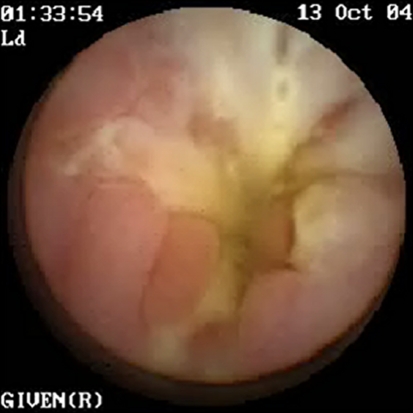
VCE image of a 14-year-old male patient with known CD. VCE was performed as small-bowel disease was suspected. Image shows severe inflammation of the small bowel with a stenosis.
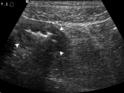
A 14-year female patient with known CD and intermittent abdominal pain. US image shows the thickened wall of the terminal ileum (arrowheads) with some infiltration of the perivisceral fat.

A 25-year-old female patient with known CD of the terminal ileum. A US image shows a large abscess (arrowheads) that was located ventrally and cranially of the bladder. B US image shows a fistula (arrowheads) that originated from the abscess.
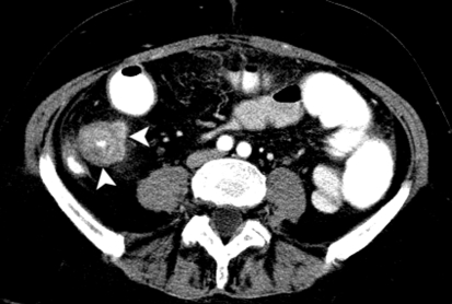
A 60-year old female patient who underwent CT enterography for suspected bowel obstruction. Just 1 month earlier at ileocolonoscopy CD of the terminal ileum was discovered; the terminal ileum was not intubated because of the stenosis. Axial image shows the severely thickened bowel wall of the ileum (arrowheads) with only a pinpoint bowel lumen remaining.
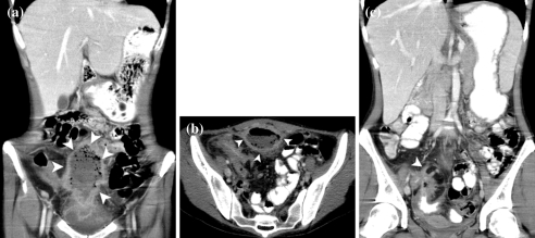
A 25-year-old female patient with known CD of the terminal ileum (same patient as pictured in Fig. 3). A CT-scan was performed to determine involvement of the small bowel. A Coronal image shows the abscess (arrowheads). B Axial image again shows the abscess again (arrowheads). C Coronal image shows the fistula (arrowhead).
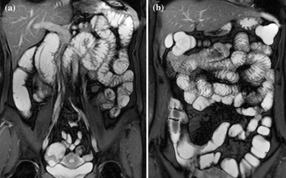
A A 38-year-old female with complaints of vomiting and an iron-deficiency anemia who was suspected of CD and underwent MRI-enteroclysis to evaluate the small bowel. Coronal TrueFISP image shows good distention of jejunal bowel loops after controlled infusion of contrast medium. B A 12-year-old male patient with known CD who underwent MR enterography for the evaluation of the small bowel. Coronal TrueFISP image shows good distention of jejunal bowel loops after oral administration of contrast medium.
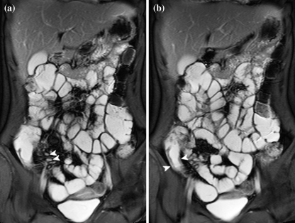
A 18-year-old female patient with known CD. MRI-enterography was performed for suspicion of active CD of the neoterminal ileum. A Coronal TrueFISp image shows enlarged mesenteric lymph nodes (arrowheads). B Coronal TrueFISP image shows thickened bowel wall of the neoterminal ileum (arrowheads).
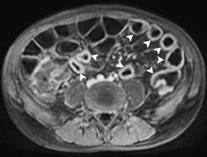
A 12-year-old male patient with known CD who underwent MR-enterography for the evaluation of the small bowel (same patient depicted in Fig. 6b). Axial T1-weighted image shows pathological enhancement of thickened small-bowel loops after administration of intravenous contrast medium (arrowheads). Approximately, 1 m of small bowel (terminal jejunum, proximal ileum) was shown to be affected.

A 25-year-old female patient with known CD of the terminal ileum (same patient as pictured in Figs. 3 and 5). A Coronal T1-weighted image clearly shows the abscess (arrowheads) that was also depicted on US and CT. B Coronal T1-weighted image showing a fistula
Similar articles
-
Inflammatory bowel disease: imaging of the pediatric patient.Semin Roentgenol. 2008 Jan;43(1):29-38. doi: 10.1053/j.ro.2007.08.005. Semin Roentgenol. 2008. PMID: 18053826 Review. No abstract available.
-
Imaging techniques in inflammatory bowel disease: recent trends, questions and answers.Gastroenterol Clin Biol. 2009 Jun;33 Suppl 3:S174-82. doi: 10.1016/S0399-8320(09)73152-5. Gastroenterol Clin Biol. 2009. PMID: 20117340
-
Imaging for Inflammatory Bowel Disease.Surg Clin North Am. 2015 Dec;95(6):1143-58, v. doi: 10.1016/j.suc.2015.07.007. Epub 2015 Sep 7. Surg Clin North Am. 2015. PMID: 26596919 Review.
-
[Bowel imaging--a reevaluation. Part 1: Conventional techniques and ultrasonography].Rofo. 2007 Jul;179(7):683-92. doi: 10.1055/s-2007-963202. Rofo. 2007. PMID: 17592807 Review. German.
-
Imaging techniques in IBD and their role in follow-up and surveillance.Nat Rev Gastroenterol Hepatol. 2014 Dec;11(12):722-36. doi: 10.1038/nrgastro.2014.144. Epub 2014 Aug 26. Nat Rev Gastroenterol Hepatol. 2014. PMID: 25157623 Review.
Cited by
-
Magnetic resonance colonography in severe attacks of ulcerative colitis.Eur Radiol. 2012 Sep;22(9):1963-71. doi: 10.1007/s00330-012-2456-8. Epub 2012 Apr 27. Eur Radiol. 2012. PMID: 22538631
-
Can diffusion weighted imaging be used as an alternative to contrast-enhanced imaging on magnetic resonance enterography for the assessment of active inflammation in Crohn disease?Medicine (Baltimore). 2020 Feb;99(8):e19202. doi: 10.1097/MD.0000000000019202. Medicine (Baltimore). 2020. PMID: 32080107 Free PMC article.
-
Evidence-based clinical practice guidelines for Crohn's disease, integrated with formal consensus of experts in Japan.J Gastroenterol. 2013 Jan;48(1):31-72. doi: 10.1007/s00535-012-0673-1. Epub 2012 Oct 23. J Gastroenterol. 2013. PMID: 23090001 Free PMC article.
-
Chronic inflammatory diseases of the bowel: diagnosis and follow-up.Pediatr Radiol. 2010 Jun;40(6):920-6. doi: 10.1007/s00247-010-1627-5. Epub 2010 Apr 30. Pediatr Radiol. 2010. PMID: 20432009 Review.
-
Contrast enhanced ultrasound: comparing a novel modality to MRI to assess for bowel disease in pediatric Crohn's patients.Transl Gastroenterol Hepatol. 2020 Apr 5;5:13. doi: 10.21037/tgh.2019.11.02. eCollection 2020. Transl Gastroenterol Hepatol. 2020. PMID: 32258517 Free PMC article.
References
-
- {'text': '', 'ref_index': 1, 'ids': [{'type': 'DOI', 'value': '10.1053/j.gastro.2004.01.063', 'is_inner': False, 'url': 'https://doi.org/10.1053/j.gastro.2004.01.063'}, {'type': 'PubMed', 'value': '15168363', 'is_inner': True, 'url': 'https://pubmed.ncbi.nlm.nih.gov/15168363/'}]}
- Loftus EV Jr (2004) Clinical epidemiology of inflammatory bowel disease: incidence, prevalence, and environmental influences. Gastroenterology 126:1504–1517 - PubMed
-
- {'text': '', 'ref_index': 1, 'ids': [{'type': 'PubMed', 'value': '11218235', 'is_inner': True, 'url': 'https://pubmed.ncbi.nlm.nih.gov/11218235/'}]}
- Farrokhyar F, Swarbrick ET, Irvine EJ (2001) A critical review of epidemiological studies in inflammatory bowel disease. Scand J Gastroenterol 36:2–15 - PubMed
-
- None
- Stenson WF (2004) Inflammatory bowel disease, Chap. 142 (on-line edition). In: Goldman L, Aussiello D, (eds). Cecil textbook of medicine, 22nd edn. Philadelphia, PA: W. B. Saunder
-
- None
- Friedman S, Blumberg RS (2005) Inflammatory bowel disease, Chap. 172 (on-line edition). In: Kasper DL, Fauci AS, Longo DL, Braunwald E, Hauser SL, Jameson JL, (eds). Harrison’s principles of internal medicine, 16th edn. New York, NY: McGraw-Hill
-
- {'text': '', 'ref_index': 1, 'ids': [{'type': 'DOI', 'value': '10.1136/gut.2005.081950a', 'is_inner': False, 'url': 'https://doi.org/10.1136/gut.2005.081950a'}, {'type': 'PMC', 'value': 'PMC1859998', 'is_inner': False, 'url': 'https://pmc.ncbi.nlm.nih.gov/articles/PMC1859998/'}, {'type': 'PubMed', 'value': '16481628', 'is_inner': True, 'url': 'https://pubmed.ncbi.nlm.nih.gov/16481628/'}]}
- Stange EF, Travis SP, Vermeire S, et al. (2006) European evidence based consensus on the diagnosis and management of Crohn’s disease: definitions and diagnosis. Gut 55(Suppl 1):i1–i15 - PMC - PubMed
Publication types
MeSH terms
Substances
LinkOut - more resources
Full Text Sources
Medical
