. 2007 Jul 9;8(1):51.
doi: 10.1186/1465-9921-8-51.

IL-13-induced proliferation of airway epithelial cells: mediation by intracellular growth factor mobilization and ADAM17

Affiliations

IL-13-induced proliferation of airway epithelial cells: mediation by intracellular growth factor mobilization and ADAM17

Brian W Booth et al. Respir Res. .

Abstract

Background: The pleiotrophic cytokine interleukin (IL)-13 features prominently in allergic and inflammatory diseases. In allergic asthma, IL-13 is well established as an inducer of airway inflammation and tissue remodeling. We demonstrated previously that IL-13 induces release of transforming growth factor-alpha (TGFalpha) from human bronchial epithelial cells, with proliferation of these cells mediated by the autocrine/paracrine action of this growth factor. TGFalpha exists as an integral membrane protein and requires proteolytic processing to its mature form, with a disintegrin and metalloproteinase (ADAM)17 responsible for this processing in a variety of tissues.

Methods: In this study, normal human bronchial epithelial (NHBE) cells grown in air/liquid interface (ALI) culture were used to examine the mechanisms whereby IL-13 induces release of TGFalpha and cellular proliferation. Inhibitors and antisense RNA were used to examine the role of ADAM17 in these processes, while IL-13-induced changes in the intracellular expression of TGFalpha and ADAM17 were visualized by confocal microscopy.

Results: IL-13 was found to induce proliferation of NHBE cells, and release of TGFalpha, in an ADAM17-dependent manner; however, this IL-13-induced proliferation did not appear to result solely from ADAM17 activation. Rather, IL-13 induced a change in the location of TGFalpha expression from intracellular to apical regions of the NHBE cells. The apical region was also found to be a site of significant ADAM17 expression, even prior to IL-13 stimulation.

Conclusion: Results from this study indicate that ADAM17 mediates IL-13-induced proliferation and TGFalpha shedding in NHBE cells. Furthermore, they provide the first example wherein a cytokine (IL-13) induces a change in the intracellular expression pattern of a growth factor, apparently inducing redistribution of intracellular stores of TGFalpha to the apical region of NHBE cells where expression of ADAM17 is prominent. Thus, IL-13-induced, ADAM17-mediated release of TGFalpha, and subsequent epithelial cell proliferation, could contribute to the epithelial hypertrophy, as well as other features, associated with airway remodeling in allergic asthma.


Figures
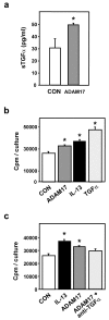
Figure 1
Figure 1
ADAM17-induced proliferation is mediated by TGFα. a) NHBE cells were treated with rhADAM17 (10 ng/ml) for 1 hr after which surrounding medium was analyzed for the presence of TGFα by ELISA (n = 3, *p < 0.05 vs. CON). b) NHBE cells were treated with rhADAM17 (10 ng/ml), IL-13 (10 ng/ml), or TGFα (5 ng/ml) for 24 hrs. [3H]-thymidine incorporation was used as a measure of proliferation (n = 6, *p < 0.05 vs. CON). c) NHBE cells were treated with IL-13 (10 ng/ml), ADAM17 (10 ng/ml) or ADAM17 plus neutralizing anti-TGFα antibody (0.2 μg/ml) for 24 hrs, with [3H]-thymidine incorporation used as a measure of proliferation (n = 6, *p < 0.05 vs. CON).
Figure 2
Figure 2
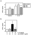
Inhibitors of ADAM17 attenuate IL-13-induced shedding of TGFα. NHBE cells were exposed to control media, inhibitors of ADAM17, IL-13 or IL-13 plus inhibitors for 1 hr. a) NHBE cells were exposed to either control media (no inhibitor), TIMP-1 or TIMP-3 (both at 2 μg/ml) for 30 min prior to treatment with IL-13 (10 ng/ml) or control media. The inhibitors were also included during the treatment period. After the 1 hr treatment, supernatants were examined for TGFα shedding via ELISA (n = 4, *p < 0.05 vs. corresponding control, †p < 0.05 vs. IL-13 alone). Light gray bars = TIMP-1; Dark gray bars = TIMP-3. b) NHBE cells were exposed to control media, anti-ADAM17 antibodies, IL-13, or IL-13 plus anti-ADAM17 for 1 hr. Supernatants were then examined for shed TGFα via ELISA (n = 6, *p < 0.05 vs. media control, †p < 0.05 vs. IL-13 alone).
Figure 3
Figure 3
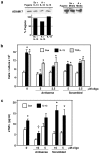
Blocking endogenous ADAM17 inhibits IL-13-induced effects. Antisense oligonucleotides directed against ADAM17 (antisense), or corresponding scrambled oligonucleotides (scrambled), were added to NHBE cell cultures for 2 days. Cultures containing no oligonucleotides received the transfection reagent (FuGene6) during this time. On the third day, cells were exposed to control media, IL-13 (10 ng/ml), or TGFα (5 ng/ml), with or without the addition of the scrambled or antisense oligonucleotides for 24 hrs. a) Total protein was extracted from a single culture from each treatment group and from the FuGene-only control group. ADAM17 was immunoprecipitated from these extracts and subjected to Western analysis (A = antisense oligonucleotides; Sc = scrambled oligonucleotides; 10 μM). The percentage of ADAM17 in experimental cultures compared to a FuGene-only exposed culture (Fugene) was determined by densitometry as indicated (left panel). The right panel was overexposed to verify the location of the two, expected ADAM17 bands. Both blots reveal decreased expression of ADAM17 in the two cultures exposed to antisense oligonucleotides. b) Cell number was determined as a measure of proliferation (n = 6, *p < 0.05 compared to appropriate control, †p < 0.01 compared to appropriate IL-13-treated, scrambled oligo sample), and c) the amount of TGFα in the supernatant was quantified via ELISA (n = 4, *p < 0.05 compared to appropriate control, †p < 0.01 compared to appropriate treated, scrambled oligo sample).
Figure 4
Figure 4
IL-13-induced effects are not due solely to activation of ADAM17. a) NHBE cells were exposed to IL-13 (10 ng/ml) or control media for 4 or 24 hrs, and steady-state mRNA levels of ADAM17 and β-actin determined via RT-PCR. Ethidium bromide-stained gels of PCR products are shown. b) NHBE cells were treated with control media or IL-13 for the times indicated. Total protein from these cells was examined for ADAM17 expression via Western blot. Membranes were chemically stripped and rehybridized to detect β-actin as a control for equal protein loading. c) NHBE cells were treated with control media, IL-13, or PMA (10 nM) for 1 hr and the supernatants examined for soluble TGFα via ELISA (n = 4, *p < 0.05 compared to control). d) NHBE cells were treated for 24 hrs with control media, IL-13, or PMA (10 nM), and [3H]-thymidine incorporation determined as a measure of proliferation (n = 6, *p < 0.05 compared to control). e) Secretion of IL-8 from NHBE cells was examined by ELISA following 1 hr exposure to control media, IL-13, or PMA (10 nM) (n = 6, *p < 0.05 compared to control).
Figure 5
Figure 5
TGFα and ADAM17 expression patterns are consistent with IL-13-induced movement of TGFα. Confocal microscopy was used to determine the cellular distribution of TGFα and ADAM17 in NHBE cells following stimulation with IL-13 for various lengths of time. Representative images from cultures of NHBE cells treated with media only (control) or IL-13 (10 ng/ml) for 15 or 30 min are shown. NHBE cultures were imaged in Z-stack mode from the basal to the apical boundaries of the cells. Images shown are x-y planes (large squares) halfway between the basal-most and the apical-most images, bordered by corresponding y-z planes (shown at right of x-y plane) and x-z planes (shown at bottom of x-y plane). The y-z and x-z plane images are from the sites indicated by the white arrows at the bottom and the right of the x-y plane images, respectively. a → b denotes the apical (a) to basal (b) direction as it relates to the x-z and y-z planes. TGFα (red) and ADAM17 (green); scale bars represent 10 μm.
Figure 6
Figure 6
Summary of TGFα and ADAM17 expression patterns induced by IL-13. a) Confocal images (y-z plane; apical to basal cross-section) of NHBE cells exposed for 60 min to media alone (control) or IL-13 (10 ng/ml). See Additional files 1 and 2 for movies of Z-stack images (basal to apical) taken from a control culture and an IL-13-treated culture, respectively, at this time point. TGFα (red) and ADAM17 (green); scale bars represent 10 μm. b) Illustration summarizing expression patterns of TGFα and ADAM17 observed via confocal microscopy in IL-13-treated NHBE cells at the times indicated. Colors represent TGFα (red) and ADAM17 (green).

Similar articles

Cited by

References

    1. McDonald DM. Angiogenesis and remodeling of airway vasculature in chronic inflammation. Am J Respir Crit Care Med. 2001;164:S39–45. - PubMed
    1. Fowlkes JL, Winkler MK. Exploring the interface between metallo-proteinase activity and growth factor and cytokine bioavailability. Cytokine Growth Factor Rev. 2002;13:277–287. doi: 10.1016/S1359-6101(02)00005-9. - DOI - PubMed
    1. Cohn L, Elias JA, Chupp GL. Asthma: mechanisms of disease persistence and progression. Annu Rev Immunol. 2004;22:789–815. doi: 10.1146/annurev.immunol.22.012703.104716. - DOI - PubMed
    1. Chung KF. The role of airway smooth muscle in the pathogenesis of airway wall remodeling in chronic obstructive pulmonary disease. Proc Am Thorac Soc. 2005;2:347–354. doi: 10.1513/pats.200504-028SR. - DOI - PMC - PubMed
    1. Podolsky DK. Mucosal immunity and inflammation. V. Innate mechanisms of mucosal defense and repair: the best offense is a good defense. Am J Physiol. 1999;277:G495–G499. - PubMed

Publication types

MeSH terms

LinkOut - more resources