. 2007 Jul 9:2:14.
doi: 10.1186/1750-1326-2-14.

ApoER2 expression increases Abeta production while decreasing Amyloid Precursor Protein (APP) endocytosis: Possible role in the partitioning of APP into lipid rafts and in the regulation of gamma-secretase activity

Affiliations

ApoER2 expression increases Abeta production while decreasing Amyloid Precursor Protein (APP) endocytosis: Possible role in the partitioning of APP into lipid rafts and in the regulation of gamma-secretase activity

Rodrigo A Fuentealba et al. Mol Neurodegener. .

Abstract

Background: The generation of the amyloid-beta peptide (Abeta) through the proteolytic processing of the amyloid precursor protein (APP) is a central event in the pathogenesis of Alzheimer's disease (AD). Recent studies highlight APP endocytosis and localization to lipid rafts as important events favoring amyloidogenic processing. However, the precise mechanisms underlying these events are poorly understood. ApoER2 is a member of the low density lipoprotein receptor (LDL-R) family exhibiting slow endocytosis rate and a significant association with lipid rafts. Despite the important neurophysiological roles described for ApoER2, little is known regarding how ApoER2 regulates APP trafficking and processing.

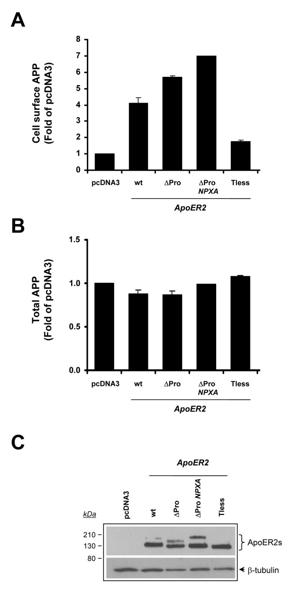
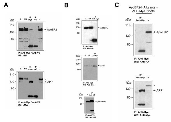
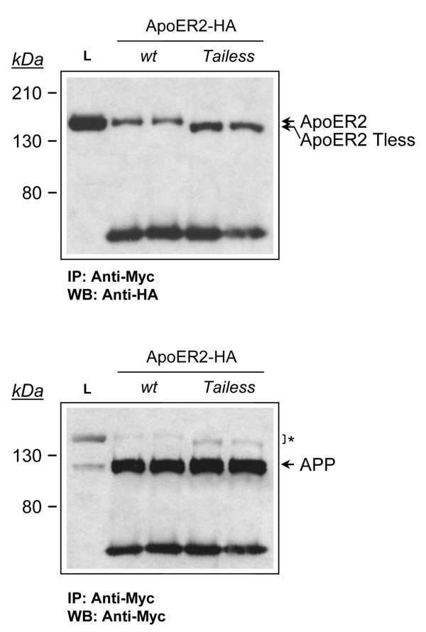
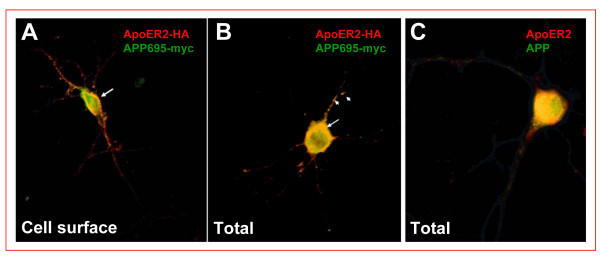
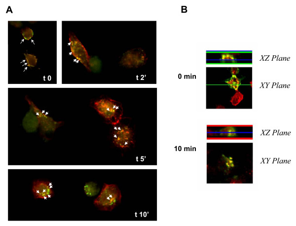
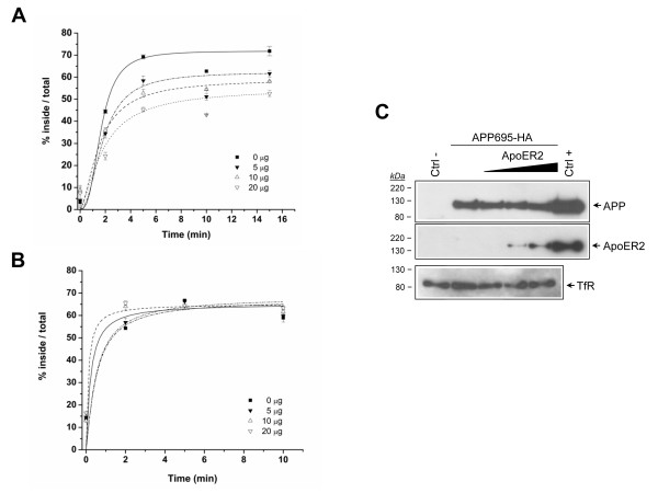
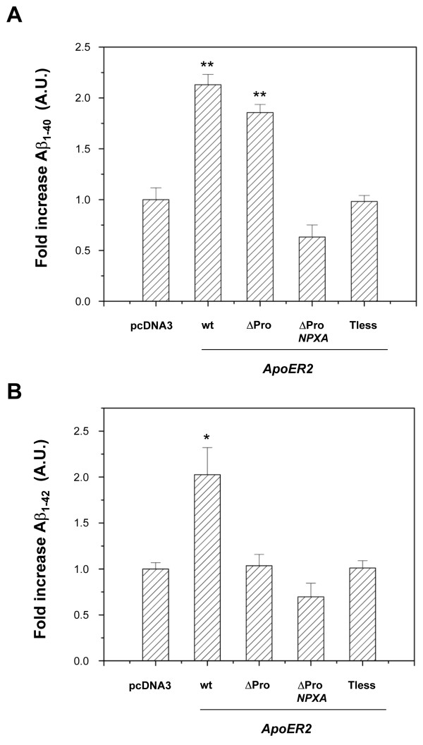
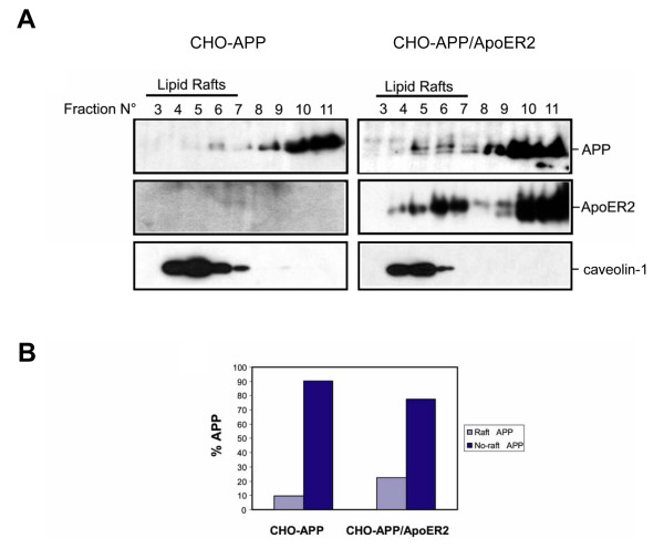
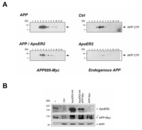
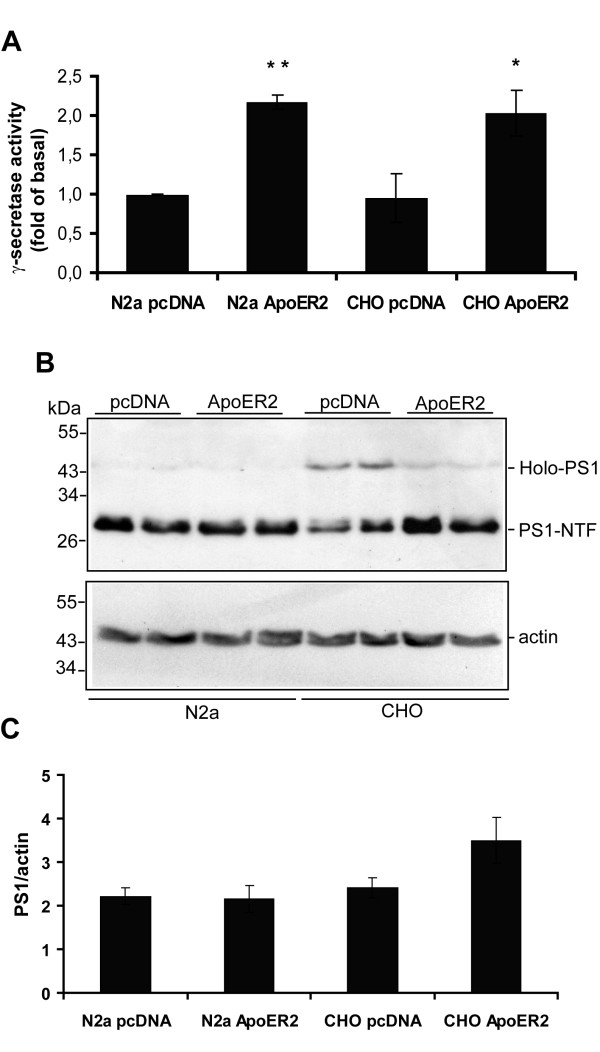
Results: Here, we demonstrate that ApoER2 physically interacts and co-localizes with APP. Remarkably, we found that ApoER2 increases cell surface APP levels and APP association with lipid rafts. The increase of cell surface APP requires the presence of ApoER2 cytoplasmic domain and is a result of decreased APP internalization rate. Unexpectedly, ApoER2 expression correlated with a significant increase in Abeta production and reduced levels of APP-CTFs. The increased Abeta production was dependent on the integrity of the NPxY endocytosis motif of ApoER2. We also found that expression of ApoER2 increased APP association with lipid rafts and increased gamma-secretase activity, both of which might contribute to increased Abeta production.

Conclusion: These findings show that ApoER2 negatively affects APP internalization. However, ApoER2 expression stimulates Abeta production by shifting the proportion of APP from the non-rafts to the raft membrane domains, thereby promoting beta-secretase and gamma-secretase mediated amyloidogenic processing and also by incrementing the activity of gamma-secretase.


Figures

Figure 1
Figure 1
ApoER2 increases cell surface APP levels but not total APP. Cell surface APP (A) and total cellular APP (B) were assessed by FACS analysis. LRP1-null CHO cells stably transfected with pcDNA3 vector, ApoER2, ApoER2ΔPro, ApoER2ΔPro-NPXA, or ApoER2-Tless were treated with or without saponin and then labeled with anti-APP antibody. Primary antibody was detected with PE-conjugated goat anti-mouse IgG. Values are the average of triplicate determinations. Error bars indicate S.E. C, lysates were prepared from CHO-pcDNA3 and ApoER2-expressing cells and equal amounts of protein were subjected to 10% SDS-PAGE. Blots were probed with anti-HA and anti-β-tubulin antibodies.
Figure 2
Figure 2
ApoER2 interacts with APP in N2a cells. (A), co-immunoprecipitation of ApoER2 with APP. Cell extracts were prepared from ApoER2-expressing cells transiently transfected with APP695-Myc. Extracts were immunoprecipitated with anti-Myc or with the unrelated, anti-V5 antibody and probed for ApoER2 and APP with the anti-HA and anti-Myc antibodies, respectively. L, input lysate. NB, not bound. (B), cell extracts were prepared from untransfected ApoER2-expressing cells and processed as in A. anti-Myc antibody does not immunoprecipitate ApoER2 by itself. Middle panel, anti-Myc antibody do immunoprecipitate APP695Myc in single transfected cells. Lower panel, anti-V5 antibody immunoprecipitation idoneity was confirmed by β-catenin-V5 precipitation from transfected N2a cells. (C), independent lysates from ApoER2-expressing cells and from APP695-Myc-transfected cells were combined and the co-immunoprecipitation experiment was continued with the anti-Myc antibody as in A. ApoER2-HA and APP695-Myc requires an in vivo context for interaction. Remnants of ApoER2 detection after stripping and reblot are indicated by an asterisk.
Figure 3
Figure 3
ApoER2 and APP interact independently of the cytosolic domain of ApoER2. Cell extracts were prepared from ApoER2- and ApoER2 Tailess-expressing N2a cells transiently transfected with APP695-Myc. Extracts were immunoprecipitated with anti-Myc and probed for ApoER2 and for APP with the anti-HA and anti-Myc antibodies, respectively. Remnants of ApoER2 detection after stripping and reblot are indicated by an asterisk.
Figure 4
Figure 4
ApoER2 and APP colocalization in hippocampal neurons. Primary cortical neurons were co-transfected with APP-Myc and ApoER2-HA and the receptors immunodetected in non-permeabilized (A) and permeabilized (B) cells using anti-Myc and anti-HA antibodies (C) Hippocampal primary neurons were permeabilized and labeled for total APP and ApoER2 with anti-APP(6E10) and C-terminal anti-ApoER2, respectively. APP695-Myc and ApoER2-HA partially co-localize at the soma surface (A, arrows) and within neuronal processes (arrowheads). A strong co-localization of APP and ApoER2 in a perinuclear region of permeabilized cells for both overexpressed (B) and endogenous levels (C) was evidenced.
Figure 5
Figure 5
ApoER2 and APP co-localize at the cell surface and within internalization vesicles of N2a cells. N2a cells were transiently transfected with ApoER2-HA and APP695-Myc and surface-immunolabeled with anti-HA and anti-Myc antibodies for 60 min at 4°C. The cells were then shifted to 37°C for varying periods of time, to allow internalization to occur. (A) Prior to warming (t0), ApoER2 and APP immunofluorescence was confirmed at the cell membrane (arrows). Partial co-immunolocalization (arrowheads) was detected as early as 2 min after endocytosis in punctuate structures near the cell membrane. By 10 minutes, most of the immunoreactivity was confined to deeper internalized vesicles, as visualized by confocal XZ-plane analysis (B) at t0 and t 10 min.
Figure 6
Figure 6
ApoER2 expression specifically decreases APP695HA endocytosis. Dose-response effect of ApoER2 expression on APP695HA (A) and diferric transferrin (B) endocytosis. CHO-LRP null cells transiently transfected with APP695HA alone (solid square) or along with 5 μg (solid inverted triangle), 10 μg (open triangle) or 20 μg (open inverted triangle) of an ApoER2 coding plasmid were incubated with 1 nM 125I-labeled-anti-HA IgG or 5 nM 125I-labeled-diFeTf for 1 h at 4°C and then shifted to 37°C for the indicated times. The percent of ligand internalized at each time point is equal to the amount of ligand internalized divided by the total cell-associated ligand (see "Materials and Methods") Values are the mean average of triplicate determinations with S.E. indicated by error bars. (C) Lysates were prepared from similarly transfected cells and equal amounts of protein were subjected to 10% SDS-PAGE. Blots were probed with anti-HA, anti-ApoER2, anti-actin or anti-transferrin receptor (TfR) antibodies. ApoER2 expression decreases APP695 but not transferrin receptor endocytosis rate.
Figure 7
Figure 7
Increased endogenous Aβ secretion in LRP1-null, ApoER2-expressing CHO cells. LRP1-null CHO cells stably transfected with pcDNA3 vector, ApoER2, ApoER2ΔPro, ApoER2ΔPro-NPXA, or ApoER2-Tless encoding plasmids were incubated in low serum media. After 48 h, media was analyzed for Aβ levels by ELISA. (A) Aβ40 and (B) Aβ42 values were normalized to the amount of cellular protein in the corresponding cell extracts and expressed as fold increase over pcDNA3-expressing cells. Values are the mean average of triplicate determinations with S.E. indicated by error bars.
Figure 8
Figure 8
ApoER2 increases full length APP association to lipid rafts in LRP1-null CHO cells. (A) Increased association of APP to lipid rafts in ApoER2-expressing cells. LRP1-null CHO cells were transiently transfected with pcDNA3 (control) and APP695-Myc (APP) or APP695-Myc along with ApoER2-HA, treated with 10 μM DAPT for 16 h and lipid rafts were isolated in Lubrol 0.5% – containing lysis buffer in a sucrose gradient. After concentration of sucrose gradients fractions, proteins were subjected to 6% SDS-PAGE, blotted and probed with anti-APP and anti-ApoER2 antibodies. To demonstrate that ApoER2 expression does not disrupts lipid rafts formation in APP expressing cells, 1/16 sucrose gradients fractions were subjected to 12.5% SDS-PAGE and probed with anti-caveolin antibody. ApoER2 increases lipid raft association of full length APP. (B), densitometric analysis of a representative Western blot experiment of (A).
Figure 9
Figure 9
ApoER2 expression decreases APP-CTFs in DAPT-treated LRP1-null CHO cells. (A) ApoER2 decreases both endogenous hamster APP-CTFs and human APP695-CTFs. LRP1-null CHO cells were transiently transfected with pcDNA (control), APP695-Myc (APP) or APP695-Myc along with ApoER2-HA (APP/ApoER2). Cells were then treated with DAPT 10 μM and protein association to lipid rafts was analyzed as in Figure 8. High sensitivity films were used for endogenous APP-CTFs detection. (B), Lubrol-extracted proteins from lysates obtained in (A) were subjected to 10% SDS-PAGE. Blots were probed with anti-ApoER2, anti-APP and anti-actin or antibodies. Similar APP protein levels were detected in APP695-Myc and in APP695-Myc + ApoER2-HA transfected cells. Remnants of ApoER2 detection after stripping and reblot are indicated by an asterisk.
Figure 10
Figure 10
ApoER2 expression enhances γ-secretase activity. N2a and CHO LRP1-null cells expressing full length ApoER2 or transfected with pcDNA3 alone were lysed in CHAPSO containing buffer. (A), Measurement of γ-secretase activity using a fluorogenic substrate assay, which is based on the secretase-dependent cleavage of a γ-secretase-specific substrate conjugated with a fluorescent molecule. Data represent mean ± S.E.M of three experiments in duplicated, presented as fold values of the basal activity. * P < 0.05, ** P < 0.01. (B), 50 μg of protein/lane were subjected to 12% SDS-PAGE and endogenous PS1 levels were determined by Western blot. In N2a cells almost all PS1 is detected as PS1-NTF but in CHO LRP1-null cells a significant amount of Holo-PS1 is also detected. ApoER2 expression increases PS1-NTF and decreases Holo-PS1 compared to control CHO LRP1-null cells. The expression of actin was determined in the same blot as a loading control. (C), densitometric analysis of Western blots as in (B) confirms that total PS1 levels were slightly increased in CHO-ApoER2 cells compared to the pcDNA3 cells. However, a further increase in the PS1-NTF fraction is clearly observed in the Western blot.

Similar articles

Cited by

References

    1. Selkoe DJ. Alzheimer's disease: genes, proteins, and therapy. Physiol Rev. 2001;81:741–766. - PubMed
    1. Howlett DR, Jennings KH, Lee DC, Clark MS, Brown F, Wetzel R, Wood SJ, Camilleri P, Roberts GW. Aggregation state and neurotoxic properties of Alzheimer beta-amyloid peptide. Neurodegeneration. 1995;4:23–32. doi: 10.1006/neur.1995.0003. - DOI - PubMed
    1. McGowan E, Pickford F, Kim J, Onstead L, Eriksen J, Yu C, Skipper L, Murphy MP, Beard J, Das P, Jansen K, Delucia M, Lin WL, Dolios G, Wang R, Eckman CB, Dickson DW, Hutton M, Hardy J, Golde T. Abeta42 is essential for parenchymal and vascular amyloid deposition in mice. Neuron. 2005;47:191–199. doi: 10.1016/j.neuron.2005.06.030. - DOI - PMC - PubMed
    1. Lambert MP, Barlow AK, Chromy BA, Edwards C, Freed R, Liosatos M, Morgan TE, Rozovsky I, Trommer B, Viola KL, Wals P, Zhang C, Finch CE, Krafft GA, Klein WL. Diffusible, nonfibrillar ligands derived from Abeta1-42 are potent central nervous system neurotoxins. Proc Natl Acad Sci U S A. 1998;95:6448–6453. doi: 10.1073/pnas.95.11.6448. - DOI - PMC - PubMed
    1. Lacor PN, Buniel MC, Chang L, Fernandez SJ, Gong Y, Viola KL, Lambert MP, Velasco PT, Bigio EH, Finch CE, Krafft GA, Klein WL. Synaptic targeting by Alzheimer's-related amyloid beta oligomers. J Neurosci. 2004;24:10191–10200. doi: 10.1523/JNEUROSCI.3432-04.2004. - DOI - PMC - PubMed

LinkOut - more resources