

Insulin receptor substrate (IRS)-1 regulates murine embryonic stem (mES) cells self-renewal
- PMID: 17620314
- PMCID: PMC3760688
- DOI: 10.1002/jcp.21185
Insulin receptor substrate (IRS)-1 regulates murine embryonic stem (mES) cells self-renewal
Abstract
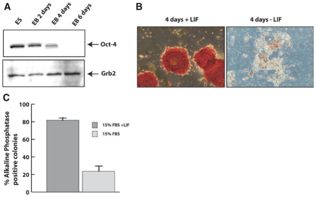
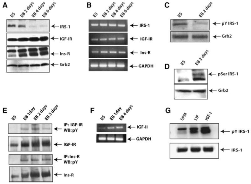
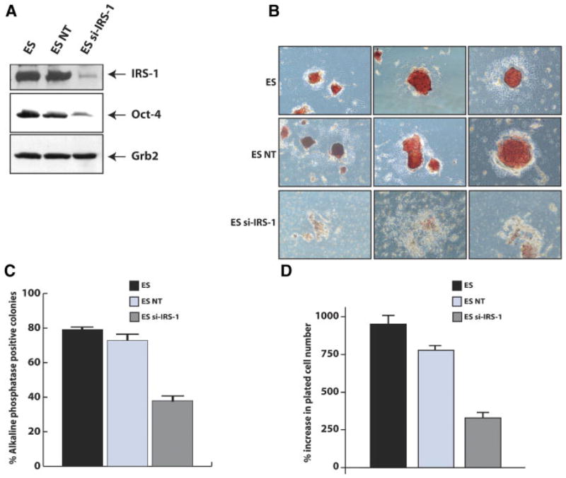
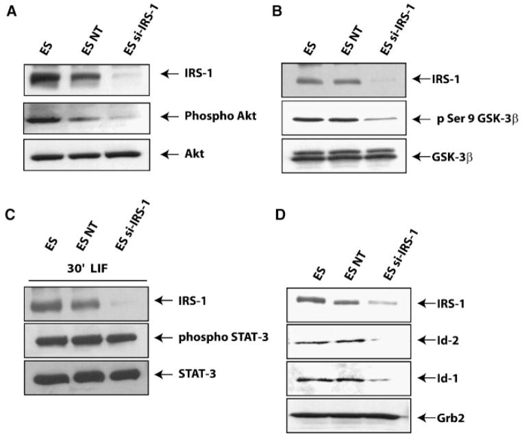
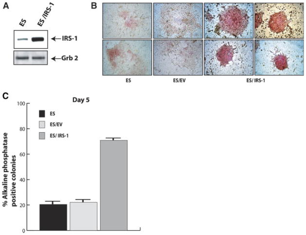
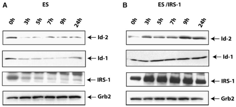
Mouse embryonic stem (mES) cells are pluripotent cells that can be propagated in vitro with leukemia inhibitory factor (LIF) and serum. Intracellular signaling by LIF is principally mediated by activation of STAT-3, although additional pathways for self-renewal have been described. Here, we identified a novel role for Insulin receptor substrate-1 (IRS-1) as a critical factor in mES cells self-renewal and differentiation. IRS-1 is expressed and tyrosyl phosphorylated during mES cells self-renewal. Differentiation of mES cells, by LIF withdrawal, is associated with a marked reduction in IRS-1 expression. Targeting of IRS-1 by si-IRS-1 results in a severe reduction of Oct-4 protein expression and alkaline phosphatase activity, markers of undifferentiated mES cells. IRS-1 targeting does not interfere with LIF-induced STAT-3 phosphorylation, but negatively affects protein kinase B (PKB/AKT) and glycogen synthase kinase-3 (GSK-3beta) phosphorylation, which are downstream effectors of the LIF-mediated PI3K signaling cascade. Targeting of IRS-1 also results in a marked down regulation of Id-1 and Id-2 proteins expression, which are important components for self-renewal of ES cells. Conversely, over expression of IRS-1 inhibits mES cell differentiation. Taken together, these results suggest that expression and activity of IRS-1 are critical to the maintenance of the self-renewal program in mES cells.
Figures

Similar articles
-
STAT3-dependent mouse embryonic stem cell differentiation into cardiomyocytes: analysis of molecular signaling and therapeutic efficacy of cardiomyocyte precommitted mES transplantation in a mouse model of myocardial infarction.Circ Res. 2007 Oct 26;101(9):910-8. doi: 10.1161/CIRCRESAHA.107.156786. Epub 2007 Sep 6. Circ Res. 2007. PMID: 17823373
-
A novel signaling by vitamin A/retinol promotes self renewal of mouse embryonic stem cells by activating PI3K/Akt signaling pathway via insulin-like growth factor-1 receptor.Stem Cells. 2010 Jan;28(1):57-63. doi: 10.1002/stem.251. Stem Cells. 2010. PMID: 19890980
-
Integrins regulate mouse embryonic stem cell self-renewal.Stem Cells. 2007 Dec;25(12):3005-15. doi: 10.1634/stemcells.2007-0103. Epub 2007 Aug 23. Stem Cells. 2007. PMID: 17717067
-
[Signaling pathways regulating self-renewal of mouse embryonic stem cells--review].Zhongguo Shi Yan Xue Ye Xue Za Zhi. 2006 Dec;14(6):1248-52. Zhongguo Shi Yan Xue Ye Xue Za Zhi. 2006. PMID: 17204204 Review. Chinese.
-
Cytokine signalling in embryonic stem cells.APMIS. 2005 Nov-Dec;113(11-12):756-72. doi: 10.1111/j.1600-0463.2005.apm_391.x. APMIS. 2005. PMID: 16480448 Review.
Cited by
-
Insulin receptor-mediated signaling regulates pluripotency markers and lineage differentiation.Mol Metab. 2018 Dec;18:153-163. doi: 10.1016/j.molmet.2018.09.003. Epub 2018 Sep 19. Mol Metab. 2018. PMID: 30316806 Free PMC article.
-
Expression and function of the insulin receptor substrate proteins in cancer.Cell Commun Signal. 2009 Jun 17;7:14. doi: 10.1186/1478-811X-7-14. Cell Commun Signal. 2009. PMID: 19534786 Free PMC article.
-
Regulation of embryonic stem cell self-renewal and pluripotency by leukaemia inhibitory factor.Biochem J. 2011 Aug 15;438(1):11-23. doi: 10.1042/BJ20102152. Biochem J. 2011. PMID: 21793804 Free PMC article. Review.
-
IGF1 regulates RUNX1 expression via IRS1/2: Implications for antler chondrocyte differentiation.Cell Cycle. 2017 Mar 19;16(6):522-532. doi: 10.1080/15384101.2016.1274471. Epub 2017 Jan 5. Cell Cycle. 2017. PMID: 28055425 Free PMC article.
-
The emerging role of insulin and insulin-like growth factor signaling in cancer stem cells.Front Endocrinol (Lausanne). 2014 Feb 4;5:10. doi: 10.3389/fendo.2014.00010. eCollection 2014. Front Endocrinol (Lausanne). 2014. PMID: 24550888 Free PMC article. Review.
References
-
- Araki E, Lipes MA, Patti ME, Bruning JC, Haag B, III, Johnson RS, Kahn CR. Alternative pathway of insulin signalling in mice with targeted disruption of the IRS-1 gene. Nature. 1994;372:186–190. - PubMed
-
- Argetsinger LS, Hsu GW, Myers MG, Jr, Billestrup N, White MF, Carter-Su C. Growth hormone, interferon-gamma, and leukemia inhibitory factor promoted tyrosyl phosphorylation of insulin receptor substrate-1. J Biol Chem. 1995;270:14685–14692. - PubMed
-
- Baserga R. The contradictions of the insulin-like growth factor 1 receptor. Oncogene. 2000;19:5574–5581. - PubMed
-
- Baserga R. The insulin-like growth factor-I receptor as a target for cancer therapy. Expert Opin Ther Targets. 2005;9:753–768. - PubMed
-
- Belletti B, Prisco M, Morrione A, Valentinis B, Navarro M, Baserga R. Regulation of Id2 gene expression by the insulin-like growth factor I receptor requires signaling by phosphatidylinositol 3-kinase. J Biol Chem. 2001;276:13867–13874. - PubMed
Publication types
MeSH terms
Substances
Grants and funding
LinkOut - more resources
Full Text Sources
Miscellaneous
