. 2007 Oct;75(10):5059-67.
doi: 10.1128/IAI.00153-07. Epub 2007 Jul 9.

Slc11a1, formerly Nramp1, is expressed in dendritic cells and influences major histocompatibility complex class II expression and antigen-presenting cell function

Affiliations

Slc11a1, formerly Nramp1, is expressed in dendritic cells and influences major histocompatibility complex class II expression and antigen-presenting cell function

Carmel B Stober et al. Infect Immun. 2007 Oct.

Abstract

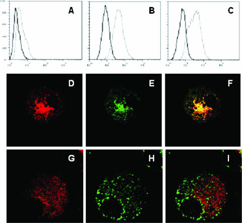
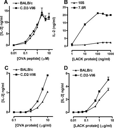
Solute carrier family 11 member a1 (Slc11a1; formerly Nramp1) encodes a late endosomal/lysosomal protein/divalent cation transporter that regulates iron homeostasis in macrophages. During macrophage activation, Slc11a1 has multiple pleiotropic effects on gene regulation and function, including gamma interferon-induced class II expression and antigen-presenting cell function. The wild-type allele at Slc11a1 has been associated with a bias in Th1 cell function in vivo, which is beneficial in resistance to infection against intracellular macrophage pathogens but detrimental in contributing to development of type 1 diabetes. The extent to which this depends on macrophage versus dendritic cell (DC) function is not known. Here we show that Slc11a1 is expressed in late endosomes and/or lysosomes of CD11c(+) DCs. DCs from mutant and congenic wild-type mice upregulate interleukin-12 (IL-12) and IL-10 mRNA in response to lipopolysaccharide (LPS) stimulation, but the ratio of IL-10 to IL-12 is higher in unstimulated DCs and DCs stimulated for 15 h with LPS from mutant mice than from wild-type mice. DCs from wild-type mice upregulate major histocompatibility complex class II in response to LPS more efficiently than DCs from mutant mice. Unstimulated DCs from wild-type and mutant mice present ovalbumin (OVA) peptide with an efficiency equivalent to that of an OVA-specific CD4 T-cell line, but DCs from wild-type mice are more efficient at processing and presenting OVA or Leishmania activator of cell kinase (LACK) protein to OVA- and LACK-specific T cells. These data indicate that wild-type Slc11a1 expressed in DCs may play a role both in determining resistance to infectious disease and in susceptibility to autoimmune disease such as type 1 diabetes.


Figures

FIG. 1.
FIG. 1.
Relative expression of Slc11a1 mRNA RAW264.7 macrophages stably transfected with wild-type (7.5R) or mutant (10S) Slc11a1, unstimulated (U/S) or stimulated with 10 U of IFN-γ/ml for 15 or 24 h (A) and in CD11c+ DCs derived from wild-type C.D2-Vil6 bone marrow, unstimulated or stimulated with 20 U of IFN-γ or 1 μg of LPS/ml for 24 h/ml (B); CD11c+ DCs derived from bone marrow from wild-type (C.D2-Vil6) and mutant (BALB/c) congenic mice on a BALB genetic background, unstimulated or stimulated with 1 μg of LPS/ml for 15, 24, or 36 h (C); and CD11c+ DCs derived from bone marrow from wild-type (B10.L-Lsh) and mutant (B10) congenic mice on a B10 genetic backgrounds, unstimulated or stimulated with 1 μg of LPS/ml for 15, 24, or 36 h (D). RNA was isolated and Slc11a1 expression was determined by using TaqMan quantitative RT-PCR. Relative quantification of signal per cell was determined by subtracting the CT for the target gene from the CT for GAPDH. Relative expression, measured as 2−ΔΔCT, is compared against an internal control (unstimulated 10S = 1 in panel A; unstimulated C.D2-Vil-6 = 1 in panel B; unstimulated C.D2-Vil6 DCs = 1 for panels C and D). All bars on the graphs show the standard error of the mean (some are too small to be visible). None of the differences between DCs from congenic pairs of mutant versus wild-type mice in panels C and D were significant (P > 0.05). Similar results were obtained in two independent experiments for the comparisons in panels C and D.
FIG. 2.
FIG. 2.
Slc11a1 protein expression in CD11c+ DCs from wild-type B10.L-Lsh mice. Slc11a1 expression was determined by using FACScan (A to C) and confocal microscopy (D to I) using an anti-N-terminal anti-Slc11a1 polyclonal antibody. FACScan profiles compare the Slc11a1-negative WEH231 B-cell line (A) with Slc11a1-positive bone marrow-derived macrophages (B) and CD11c+ DCs (C). The isotype control antibody is shown with the black line; anti-Slc11a1 is shown with the gray line. The x axis shows the mean fluorescence intensity for Slc11a1; the y axis cell shows the numbers of cells. Confocal microscopy shows that Slc11a1 protein (D and G) colocalizes with the late endosomal/lysosomal marker Lamp1 (E; Slc11a1 and Lamp1 merged in panel F) but not the early endosomal marker EEA1 (H; Slc11a1 and EEA1 merged in panel I). Similar results were obtained in three independent experiments for FACScan studies and in two independent experiments for confocal studies.
FIG. 3.
FIG. 3.
IL-12 (A and B) and IL-10 (C and D) mRNA expression in CD11c+ DCs from wild-type (C.D2-Vil6; B10.L-Lsh) and mutant (BALB/c; B10) congenic mice on BALB (A and C) and B10 (B and D) genetic backgrounds. RNA was isolated from unstimulated (U/S) DCs and from DC stimulated for 15, 24, or 36 h with 1 μg of LPS/ml. IL-12 and IL-10 expression was determined by using TaqMan quantitative RT-PCR. Relative quantification of signal per cell was determined by subtracting the CT value for the target gene from the CT for GAPDH. Relative expression, measured as 2−ΔΔCT, is compared against an internal control (unstimulated C.D2-Vil6 DC = 1). The ratio of IL-10 to IL-12 is shown for BALB (E) and B10 (F) background mice. All bars on the graphs show the standard error of the mean (some are too small to be visible). Differences in ratios are significant at P = 0.008 for unstimulated DCs and at P = 0.0008 for DCs stimulated for 15 h in panel E and at P = 0.028 for unstimulated DCs in panel F. Similar results were obtained in three independent experiments.
FIG. 4.
FIG. 4.
MHC class II (A and C) and CD40 (B and D) protein expression in CD11c+ DCs from mutant B10 (A and B) and wild-type B10.L-Lsh (C and D) mice. Class II and CD40 expression was determined by using FACScan with rat anti-mouse I-A/I-E clone M5/114.15.2 and rat anti-mouse CD40 clone 3/23. Baseline class II or CD40 expression in unstimulated DCs is indicated by the black line; upregulated expression 48 h after LPS stimulation is indicated by the gray line. Quantitative analysis of geometric mean fluorescence over time after LPS stimulation is shown for an independent experiment examining class II expression in Table 1. Similar results were obtained in three independent experiments for MHC class II; only one experiment was performed for CD40.
FIG. 5.
FIG. 5.
Antigen-presenting cell function in CD11c+ DCs (A, C, and D) from congenic C.D2-Vil6 wild-type and BALB/c mutant (BALB/c) mice compared to Slc11a1 wild-type (7.5R) and mutant (10S) stably transfected macrophage cell lines (B). The ability of DCs to present OVA peptide 323-339 to the OVA-specific DO11.10 T-cell hybridoma (A) is compared to their ability to process and present OVA protein (C). The ability to process and present recombinant LACK protein to the LACK-specific LMR7.5 T-cell hybridoma is compared for transfected macrophages (B) and DCs (D). Similar results were obtained in two independent experiments.

Similar articles

Cited by

References

    1. Atkinson, P. G. P., and C. H. Barton. 1998. Ectopic expression of Nramp1 in COS-1 cells modulates iron accumulation. FEBS Lett. 425:239-242. - PubMed
    1. Awomoyi, A. A., A. Marchant, J. M. Howson, K. P. McAdam, J. M. Blackwell, and M. J. Newport. 2002. Interleukin-10, polymorphism in SLC11A1 (formerly NRAMP1), and susceptibility to tuberculosis. J. Infect. Dis. 186:1804-1814. - PubMed
    1. Banchereau, J., and R. M. Steinman. 1998. Dendritic cells and the control of immunity. Nature 392:245-252. - PubMed
    1. Barrera, L. F., I. Kramnik, E. Skamene, and D. Radzioch. 1997. I-A beta gene expression regulation in macrophages derived from mice susceptible or resistant to infection with M. bovis BCG. Mol. Immunol. 34:343-355. - PubMed
    1. Barton, C. H., J. K. White, T. I. A. Roach, and J. M. Blackwell. 1994. NH2-terminal sequence of macrophage-expressed natural resistance-associated macrophage protein (Nramp) encodes a proline/serine-rich putative Src homology 3-binding domain. J. Exp. Med. 179:1683-1687. - PMC - PubMed

Publication types

MeSH terms