. 2007 Aug;171(2):452-62.
doi: 10.2353/ajpath.2007.060805. Epub 2007 Jul 9.

Extracellular signal-regulated kinase activation during renal ischemia/reperfusion mediates focal adhesion dissolution and renal injury

Affiliations

Extracellular signal-regulated kinase activation during renal ischemia/reperfusion mediates focal adhesion dissolution and renal injury

Maaike Alderliesten et al. Am J Pathol. 2007 Aug.

Abstract

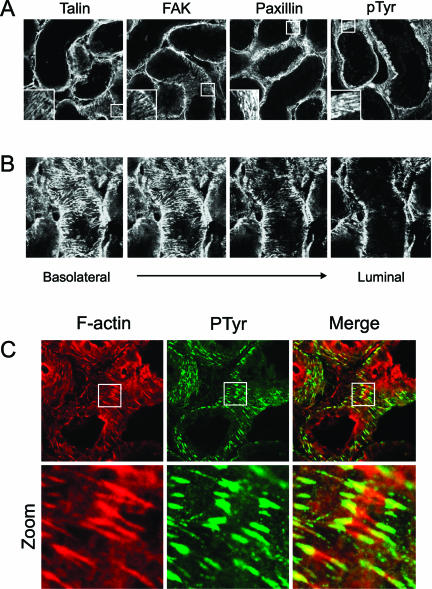
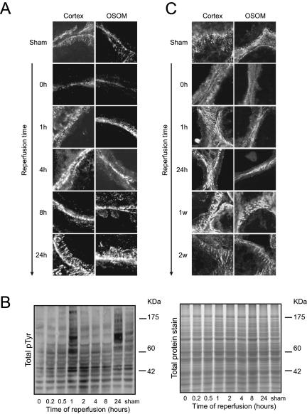
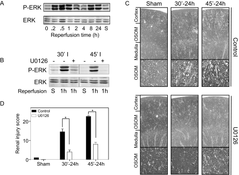
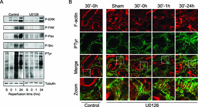
Acute renal failure due to ischemia/reperfusion involves disruption of integrin-mediated cellular adhesion and activation of the extracellular signal-regulated kinase (ERK) pathway. The dynamics of focal adhesion organization and phosphorylation during ischemia/reperfusion in relation to ERK activation are unknown. In control kidneys, protein tyrosine-rich focal adhesions, containing focal adhesion kinase, paxillin, and talin, were present at the basolateral membrane of tubular cells and colocalized with short F-actin stress fibers. Unilateral renal ischemia/reperfusion caused a reversible protein dephosphorylation and loss of focal adhesions. The focal adhesion protein phosphorylation rebounded in a biphasic manner, in association with increased focal adhesion kinase, Src, and paxillin tyrosine phosphorylation. Preceding phosphorylation of these focal adhesion proteins, reperfusion caused increased phosphorylation of ERK. The specific mitogen-activated protein kinase kinase 1/2 inhibitor U0126 prevented ERK activation and attenuated focal adhesion kinase, paxillin, and Src phosphorylation, focal adhesion restructuring, and ischemia/reperfusion-induced renal injury. We propose a model whereby ERK activation enhanced protein tyrosine phosphorylation during ischemia/reperfusion, thereby driving the dynamic dissolution and restructuring of focal adhesions and F-actin cytoskeleton during reperfusion and renal injury.


Figures

Figure 1
Figure 1
In vivo colocalization of tyrosine-phosphorylated focal adhesion proteins with F-actin stress fibers. To determine localization of FAs in the proximal tubules, frozen sections (10 μm) of control kidneys were stained for the FA proteins talin, FAK, and paxillin and for total tyrosine phosphorylation (pTyr) (A). A Z-scan was created from the basolateral site toward the lumen of the tubule to show localization of FAK (B). To indicate colocalization between pTyr containing FAs and F-actin, sections were costained for pTyr (green) and F-actin (red); colocalization is yellow (C). All sections were imaged using a confocal laser scanning microscope. Sections are representative of proximal tubules in three different rats and observed in two different stainings.
Figure 2
Figure 2
I/R caused a biphasic increase in protein tyrosine phosphorylation and restructuring of focal adhesions. Rats were subjected to 30 (A) or 45 minutes (C) of ischemia followed by reperfusion for the indicated time periods. Thereafter, frozen kidney sections (10 μm) were stained for total tyrosine phosphorylation using an anti-pTyr antibody. Both OSOM and cortex region were evaluated for differential tyrosine phosphorylation using confocal laser scanning microscopy (A and C) and Western blot analysis by staining blots with an anti-pTyr antibody (B). Total protein was measured by staining a sodium dodecyl sulfate-polyacrylamide gel electrophoresis gel with Sypro Ruby (B). Data are representative of three different rats and observed in two different stainings.
Figure 3
Figure 3
I/R induces dynamic phosphorylation of FAK, paxillin, and Src. Rats were subjected to 30 minutes of ischemia followed by reperfusion for 0 to 24 hours. Frozen sections (10 μm) were stained for FAK (green) and PY397-FAK (red) to determine differential tyrosine phosphorylation and colocalization (yellow) at FAs (A). In addition, frozen sections were prepared for Western blot analysis and stained for PY397-FAK and FAK (B), PY118-paxillin and paxillin (D), and PY576-FAK, PY861-FAK PY416-Src kinase, and PSer17/221-ERK (C). All sections were imaged using a confocal laser scanning microscope. Data are representative of proximal tubules in three different rats.
Figure 4
Figure 4
I/R-induced injury is inhibited by the ERK inhibitor U0126. Rats were pretreated with U0126 (1 mg/kg) 30 minutes before I/R for indicated time points. For untreated (A) and treated rats (B), frozen sections were prepared for Western blot analysis and stained for PSer17/221-ERK. Paraffin sections were stained for H&E to determine tubular damage (C). Sections are representative of proximal tubules in three different rats. Pictures were made using light microscopy (Leica DM6000B; magnifications, ×25 and ×100). Sections were scored double-blind and semiquantitatively to assess tubulointerstitial injury (D). Data are presented as means ± SE (n = 3 rats per group). *P < 0.05 compared with control.
Figure 5
Figure 5
U0126 prevents I/R-mediated loss of FA tyrosine phosphorylation in conjunction with maintenance of FA structure. Rats were pretreated with U0126 (1 mg/kg), and both control and U0126-treated rats were subjected to 30 minutes of ischemia followed by reperfusion for the indicated time periods. Thereafter, frozen kidney sections (10 μm) were prepared for Western blotting and stained for pTyr, P-FAK, paxillin, and Src (A) and stained with rhodamine/phalloidin for F-actin and total tyrosine phosphorylation using anti-pTyr antibody (B). Sections were evaluated for F-actin reorganization and differential pTyr using confocal laser scanning microscopy. The bottom panel indicates a zoom of the areas containing stress fibers and tyrosine-phosphorylated focal adhesions. Sections are representative of proximal tubules in three different rats.

Similar articles

Cited by

References

    1. Molina A, Ubeda M, Escribese MM, Garcia-Bermejo L, Sancho D, de Lema GP, Liano F, Cabanas C, Sanchez-Madrid F, Mampaso F. Renal ischemia/reperfusion injury: functional tissue preservation by anti-activated β1 integrin therapy. J Am Soc Nephrol. 2005;16:374–382. - PubMed
    1. Zalewska T, Makarewicz D, Janik B, Ziemka-Nalecz M. Neonatal cerebral hypoxia-ischemia: involvement of FAK-dependent pathway. Int J Dev Neurosci. 2005;23:657–662. - PubMed
    1. Zalewska T, Ziemka-Nalecz M, Domanska-Janik K. Transient forebrain ischemia effects interaction of Src FAK, and PYK2 with the NR2B subunit of N-methyl-D-aspartate receptor in gerbil hippocampus. Brain Res. 2005;1042:214–223. - PubMed
    1. Zuk A, Bonventre JV, Brown D, Matlin KS. Polarity, integrin, and extracellular matrix dynamics in the postischemic rat kidney. Am J Physiol. 1998;275:C711–C731. - PubMed
    1. Raman N, Atkinson SJ. Rho controls actin cytoskeletal assembly in renal epithelial cells during ATP depletion and recovery. Am J Physiol. 1999;276:C1312–C1324. - PubMed

Publication types

MeSH terms

LinkOut - more resources