

Osteopontin gene expression determines spontaneous metastatic performance of orthotopic human breast cancer xenografts
- PMID: 17620367
- PMCID: PMC1934534
- DOI: 10.2353/ajpath.2007.070232
Osteopontin gene expression determines spontaneous metastatic performance of orthotopic human breast cancer xenografts
Abstract
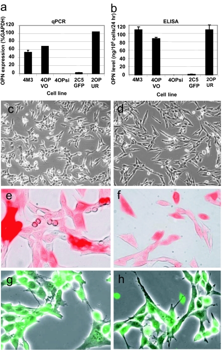
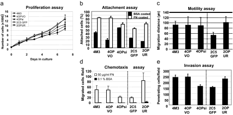
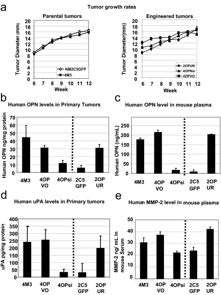
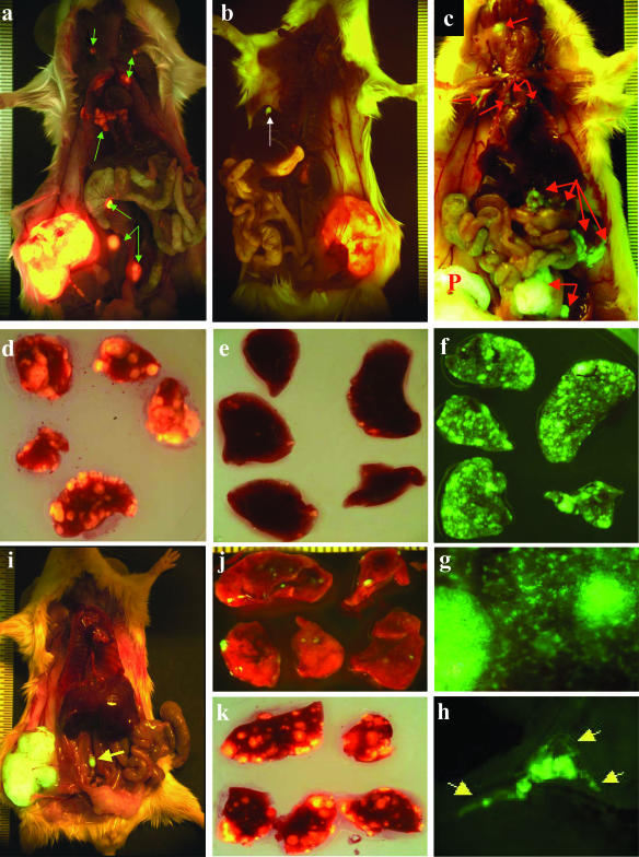
A major problem in the therapeutic management of cancer is the growth of metastases in distant organs, but the genes orchestrating the process need to be identified for the rational design of new treatment. Here, we provide decisive experimental evidence demonstrating the causal involvement of a specific gene, osteopontin (OPN), in the pathogenesis of metastasis by human breast cancer cells and implicating some of its probable partners. Stable long-term depletion, or up-regulation, of OPN gene expression in a matched, isogenic pair of human breast cancer cell lines of differing metastatic proficiency reproducibly changed their ability to colonize distant organs. OPN down-regulation was achieved by transduction of the metastatic line with a DNA construct encoding a small hairpin RNA in a vector labeled with red fluorescent protein and resulted in a marked reduction of metastatic load (P < 0.01). Up-regulation of OPN in the negligibly metastatic line, with a green fluorescent protein-marked retroviral vector containing OPN cDNA driven by a strong promoter, resulted in heavy colonization of the lungs and lymph nodes (P < 0.005). The reciprocal changes in behavior of these matched cell lines cross-corroborate each other. Concomitant changes were seen in the expression of other metastasis-related genes in both modulated lines. The data indicate that therapeutic targeting of tumor OPN molecules could reset metastatically relevant gene networks, resulting in clinical benefit.
Figures
Similar articles
-
Suppression of distant pulmonary metastasis of MDA-MB 435 human breast carcinoma established in mammary fat pads of nude mice by retroviral-mediated TIMP-2 gene transfer.J Gene Med. 2005 Feb;7(2):145-57. doi: 10.1002/jgm.645. J Gene Med. 2005. PMID: 15546163
-
Osteopontin induces increased invasiveness and plasminogen activator expression of human mammary epithelial cells.Oncogene. 1999 Jul 22;18(29):4237-46. doi: 10.1038/sj.onc.1202799. Oncogene. 1999. PMID: 10435636
-
Contrasting expression of thrombospondin-1 and osteopontin correlates with absence or presence of metastatic phenotype in an isogenic model of spontaneous human breast cancer metastasis.Clin Cancer Res. 2002 Jan;8(1):61-74. Clin Cancer Res. 2002. PMID: 11801541
-
Osteopontin overexpression in breast cancer: knowledge gained and possible implications for clinical management.J Cell Biochem. 2007 Nov 1;102(4):859-68. doi: 10.1002/jcb.21520. J Cell Biochem. 2007. PMID: 17721886 Review.
-
Osteopontin at the Crossroads of Inflammation and Tumor Progression.Mediators Inflamm. 2017;2017:4049098. doi: 10.1155/2017/4049098. Epub 2017 Jul 9. Mediators Inflamm. 2017. PMID: 28769537 Free PMC article. Review.
Cited by
-
A humanized anti-osteopontin antibody inhibits breast cancer growth and metastasis in vivo.Cancer Immunol Immunother. 2010 Mar;59(3):355-66. doi: 10.1007/s00262-009-0754-z. Epub 2009 Aug 19. Cancer Immunol Immunother. 2010. PMID: 19690854 Free PMC article.
-
Osteopontin: A Key Regulator of Tumor Progression and Immunomodulation.Cancers (Basel). 2020 Nov 15;12(11):3379. doi: 10.3390/cancers12113379. Cancers (Basel). 2020. PMID: 33203146 Free PMC article. Review.
-
Growth of v-src-transformed cells in serum-free medium through the induction of growth factors.J Cell Physiol. 2013 Jul;228(7):1482-6. doi: 10.1002/jcp.24303. J Cell Physiol. 2013. PMID: 23254450 Free PMC article.
-
Osteopontin regulates proliferation, apoptosis, and migration of murine claudin-low mammary tumor cells.BMC Cancer. 2016 Jun 10;16:359. doi: 10.1186/s12885-016-2396-9. BMC Cancer. 2016. PMID: 27282619 Free PMC article.
-
Clinical and biological implications of the tumor microenvironment.Cancer Microenviron. 2012 Aug;5(2):95-112. doi: 10.1007/s12307-012-0099-6. Cancer Microenviron. 2012. PMID: 22782446 Free PMC article.
References
-
- Wai PY, Kuo PC. The role of osteopontin in tumor metastasis. J Surg Res. 2004;121:228–241. - PubMed
-
- Weber GF. The metastasis gene osteopontin: a candidate target for cancer therapy. Biochim Biophys Acta. 2001;1552:61–85. - PubMed
-
- Ehrchen J, Heuer H, Sigmund R, Schafer MK, Bauer K. Expression and regulation of osteopontin and connective tissue growth factor transcripts in rat anterior pituitary. J Endocrinol. 2001;169:87–96. - PubMed
Publication types
MeSH terms
Substances
LinkOut - more resources
Full Text Sources
Other Literature Sources
Medical
Research Materials
