. 2007 Aug;171(2):608-16.
doi: 10.2353/ajpath.2007.070068. Epub 2007 Jul 9.

Mice deficient for the type II transmembrane serine protease, TMPRSS1/hepsin, exhibit profound hearing loss

Affiliations

Mice deficient for the type II transmembrane serine protease, TMPRSS1/hepsin, exhibit profound hearing loss

Michel Guipponi et al. Am J Pathol. 2007 Aug.

Abstract

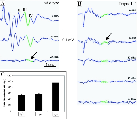
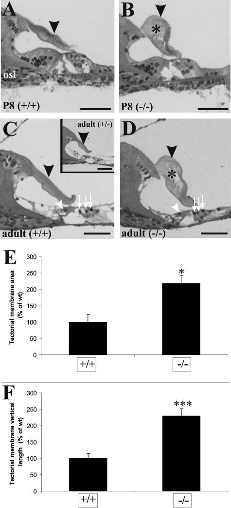
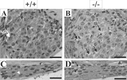
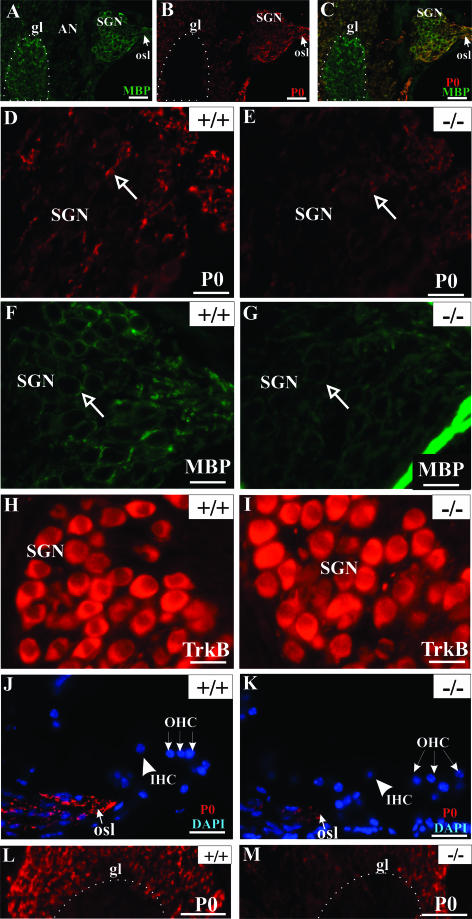
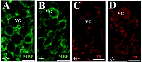
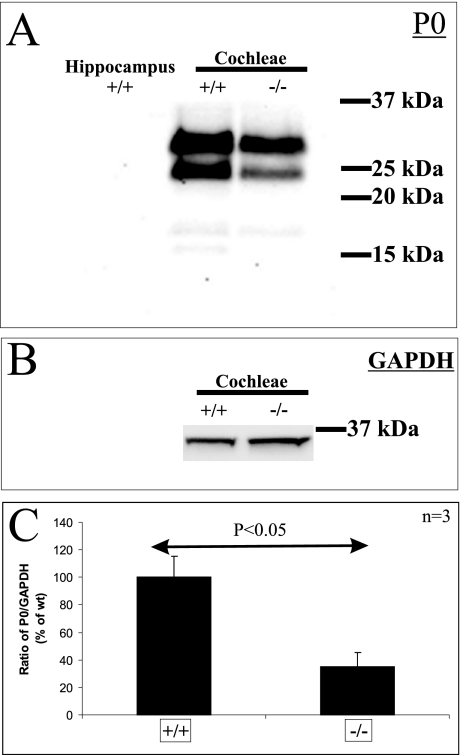
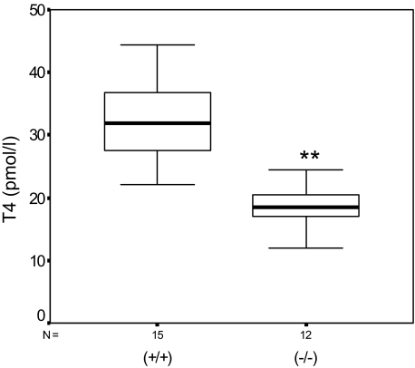
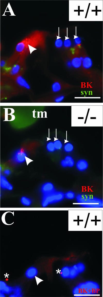
Defective proteolysis has been implicated in hearing loss through the discovery of mutations causing autosomal recessive nonsyndromic deafness in a type II transmembrane serine protease gene, TMPRSS3. To investigate their physiological function and the contribution of this family of proteases to the auditory function, we analyzed the hearing status of mice deficient for hepsin, also known as TMPRSS1. These mice exhibited profound hearing loss with elevated hearing thresholds compared with their heterozygous and wild-type littermates. Their cochleae showed abnormal tectorial membrane development, reduction in fiber compaction in the peripheral portion of the auditory nerve, and decreased expression of the myelin proteins myelin basic protein and myelin protein zero. In addition, reduced level of the large conductance voltage- and Ca(2+)-activated K(+) channel was detected in the sensory hair cells of Tmprss1-null mice. We examined thyroid hormone levels in Tmprss1-deficient mice, as similar cochlear defects have been reported in animal models of hypothyroidism, and found significantly reduced free thyroxine levels. These data show that TMPRSS1 is required for normal auditory function. Hearing impairment present in Tmprss1-null mice is characterized by a combination of various structural, cellular, and molecular abnormalities that are likely to affect different cochlear processes.


Figures

Figure 1
Figure 1
Hearing function deficit in Tmprss1−/− mice. Adult wild-type, Tmprss1 heterozygous, and Tmprss1 knockout mice were subjected to click-evoked ABR measurements. The amplitude of the response (four peaks labeled from I to IV) is measured in millivolts. The latency is shown in milliseconds. Click stimulus intensity is indicated in decibel attenuation. Maximum stimulus intensity corresponds to 0 dBA. The green portion of the graph highlights the waveform chosen to determine threshold. A: Representative ABR response of wild-type mice at 0, 20, and 40 dBA. Arrow indicates ABR threshold. B: Representative ABR response of Tmprss1−/− mice at 0, 5, 10, 15, and 20 dBA. Arrow indicates ABR threshold. C: Quantification of the average ABR thresholds of wild-type, Tmprss1 heterozygote, and Tmprss1-null mice. Graph indicates mean ± SEM.
Figure 2
Figure 2
Tectorial membrane defects in Tmprss1−/− mice. Histological analysis of cochleae from wild-type and Tmprss1−/− mice at postnatal day 8 (P8) (A and B, respectively) and adult (C and D, respectively) mice. Black arrowheads point to the tectorial membrane, and asterisks show holes found within the matrix of the membrane. Osseous spiral lamina is indicated by osl. White arrowheads show inner hair cells, and white arrows point to the outer hair cells in the organ of Corti. Scale bar = 50 μm. C, inset: Analysis of cochleae from adult Tmprss1+/− mice. E: Comparison of the cross-sectional area of the tectorial membrane in wild-type and Tmprss1−/− mice. Graph indicates mean ± SE, *P = 0.011. F: Comparison of the longest vertical distance measured between the dorsal and ventral surface of the tectorial membrane. Graph indicates mean ± SE, ***P = 0.002.
Figure 3
Figure 3
Morphological defects in the Rosenthal’s canal and osseous spiral lamina of Tmprss1−/− mice. Representative images of the Rosenthal’s canal and the osseous spiral lamina of adult wild-type (A and C, respectively) and adult Tmprss1−/− (B and D, respectively) mice. A: White arrowhead indicates spiral ganglion neuron (SGN). B: Black arrows indicate spaces between spiral ganglion neurons. C: White arrowhead indicates spindle-shaped nuclei of Schwann cells. D: Black arrows point to spaces between Schwann cells. Scale bar = 20 μm.
Figure 4
Figure 4
MBP and P0 expression are reduced in the cochleae of Tmprss1−/− mice. MBP (A) and P0 (B) expression in the auditory nerve of wild-type mice. C: Superimposition of A and B. P0, MBP and tyrosine kinase receptor B expression in the Rosenthal’s canal of adult wild-type (D, F, and H, respectively) and adult Tmprss1−/− mice (E, G, and I, respectively). D and E: Arrows indicate a spiral ganglion neuron fiber (D and E) and a spiral ganglion neuron body (F and G). The bony boundary of the Rosenthal’s canal shows strong nonspecific staining (G). P0 expression in the osseous spiral lamina and fibers projecting to auditory nerve in the brainstem of adult wild-type and adult Tmprss1−/− mice (J and K, respectively, and L and M, respectively). Scale bar = 20 μm, except in A, B, C, L, and M, where scale bar = 50 μm. Glial transition zone (gl) is approximately outlined by the white dotted line. osl, osseous spiral lamina; SGN, spiral ganglion neuron; AN, auditory nerve; IHC, inner hair cell; OHC, outer hair cell.
Figure 5
Figure 5
MBP and P0 expression in the vestibular ganglion of adult wild-type (A and C, respectively) and Tmprss1−/− mice (B and D, respectively).
Figure 6
Figure 6
Western blot quantification of the reduction of P0 expression in cochleae of Tmprss1−/− mice. A: Western blot analysis of P0 was performed on equal amount of membrane proteins isolated from hippocampus, wild-type (+/+), and Tmprss1−/− (−/−) mice cochleae. B: Western blot analysis of glyceraldehyde-3-phosphate dehydrogenase was performed on equal amounts of cytosolic proteins isolated from wild-type and Tmprss1−/− mice cochleae. C:The intensity of the 25-kd fragment was normalized to the GAPDH band in three independent experiments, and the mean ratio expressed as a relative percentage of the wild-type cohort. There was a significant reduction in this ratio in Tmprss1−/−mice, relative to wild-type mice (P= 0.024).
Figure 7
Figure 7
Tmprss1−/− mice are hypothyroidic. Box plot diagram of fT4 levels in the serum of 15 adult wild-type (+/+) and 12 Tmprss1−/− (−/−) mice. The distribution of fT4 levels between the two groups is statistically significant (**P = 2 × 10⁻⁵).
Figure 8
Figure 8
Reduced BK α-subunit expression in the organ of Corti of Tmprss1−/− mice. BK α-subunit (red) and synaptophysin (green) expression in the organ of Corti of adult wild-type (A) and age-matched Tmprss1−/− (B) mice. C: The BK α-subunit antibody was preincubated with the corresponding anti-peptide before being applied to sections. White arrowhead indicates the upper part of inner hair cells. White downward arrows point to the base of outer hair cells. Asterisks indicate background fluorescence, which does not localize within any cellular compartments. Cell nuclei are labeled with 4′,6-diamidino-2-phenylindole dihydrochloride (blue). Scale bar = 20 μm.

References

    1. Morton NE. Genetic epidemiology of hearing impairment. Ann NY Acad Sci. 1991;630:16–31. - PubMed
    1. Seidman MD, Ahmad N, Bai U. Molecular mechanisms of age-related hearing loss. Ageing Res Rev. 2002;1:331–343. - PubMed
    1. Gratton MA, Vazquez AE. Age-related hearing loss: current research. Curr Opin Otolaryngol Head Neck Surg. 2003;11:367–371. - PubMed
    1. Scott HS, Kudoh J, Wattenhofer M, Shibuya K, Berry A, Chrast R, Guipponi M, Wang J, Kawasaki K, Asakawa S, Minoshima S, Younus F, Mehdi SQ, Radhakrishna U, Papasavvas MP, Gehrig C, Rossier C, Korostishevsky M, Gal A, Shimizu N, Bonne-Tamir B, Antonarakis SE. Insertion of beta-satellite repeats identifies a transmembrane protease causing both congenital and childhood onset autosomal recessive deafness. Nat Genet. 2001;27:59–63. - PubMed
    1. Masmoudi S, Antonarakis SE, Schwede T, Ghorbel AM, Gratri M, Pappasavas MP, Drira M, Elgaied-Boulila A, Wattenhofer M, Rossier C, Scott HS, Ayadi H, Guipponi M. Novel missense mutations of TMPRSS3 in two consanguineous Tunisian families with non-syndromic autosomal recessive deafness. Hum Mutat. 2001;18:101–108. - PubMed

Publication types

MeSH terms