

Tonsil epithelial factors may influence oropharyngeal human immunodeficiency virus transmission
- PMID: 17620369
- PMCID: PMC1934526
- DOI: 10.2353/ajpath.2007.061006
Tonsil epithelial factors may influence oropharyngeal human immunodeficiency virus transmission
Abstract
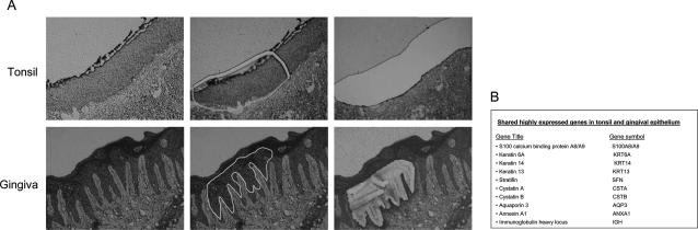
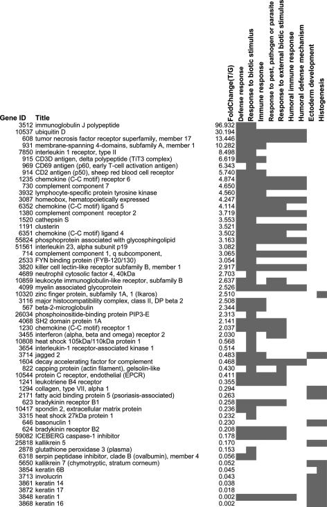
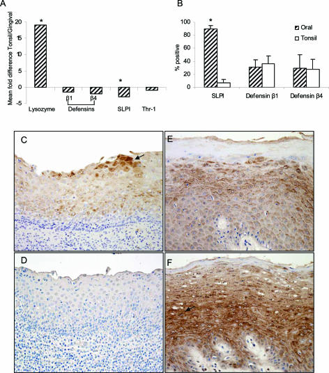
Tonsil epithelium has been implicated in human immunodeficiency virus (HIV) pathogenesis, but its role in oral transmission remains controversial. To study characteristics of this tissue, which may influence susceptibility or resistance to HIV, we performed microarray analysis of the tonsil epithelium. Our data revealed that genes related to immune functions such as antibody production and antigen processing were increasingly expressed in tonsil compared with the epithelium of another oropharyngeal site, the gingival epithelium. Importantly, tonsil epithelium highly expressed genes associated with HIV entrapment and/or transmission, including the HIV co-receptor CXCR4 and the potential HIV-binding molecules FcRgammaIII, complement receptor 2, and various complement components. Immunohistochemical staining confirmed the increased presence of CXCR4 in the tonsil epithelium compared with multiple oral epithelial sites, particularly in basal and parabasal layers. This increased expression of molecules involved in viral recognition, binding, and entry may favor virus-epithelium interactions in an environment with reduced innate antiviral mechanisms. Specifically, secretory leukocyte protease inhibitor, an innate molecule with anti-HIV activity, was minimal in the tonsil epithelium, in contrast to oral mucosa. Collectively, our data suggest that increased expression of molecules associated with HIV binding and entry coupled with decreased innate antiviral factors may render the tonsil a potential site for oral transmission.
Figures


Similar articles
-
Expression of HIV receptors, alternate receptors and co-receptors on tonsillar epithelium: implications for HIV binding and primary oral infection.Virol J. 2006 Apr 6;3:25. doi: 10.1186/1743-422X-3-25. Virol J. 2006. PMID: 16600047 Free PMC article.
-
Differential Ability of Primary HIV-1 Nef Isolates To Downregulate HIV-1 Entry Receptors.J Virol. 2015 Sep;89(18):9639-52. doi: 10.1128/JVI.01548-15. Epub 2015 Jul 15. J Virol. 2015. PMID: 26178998 Free PMC article. Clinical Trial.
-
Human Immunodeficiency Virus (HIV) and Human Cytomegalovirus (HCMV) Coinfection of Infant Tonsil Epithelium May Synergistically Promote both HIV-1 and HCMV Spread and Infection.J Virol. 2021 Aug 25;95(18):e0092121. doi: 10.1128/JVI.00921-21. Epub 2021 Aug 25. J Virol. 2021. PMID: 34232730 Free PMC article.
-
CCR5, CXCR4, and CD4 are clustered and closely apposed on microvilli of human macrophages and T cells.J Virol. 2001 Apr;75(8):3779-90. doi: 10.1128/JVI.75.8.3779-3790.2001. J Virol. 2001. PMID: 11264367 Free PMC article.
-
Molecular Pathogenesis of Human Immunodeficiency Virus-Associated Disease of Oropharyngeal Mucosal Epithelium.Biomedicines. 2023 May 14;11(5):1444. doi: 10.3390/biomedicines11051444. Biomedicines. 2023. PMID: 37239115 Free PMC article. Review.
Cited by
-
Human beta-defensins 2 and -3 cointernalize with human immunodeficiency virus via heparan sulfate proteoglycans and reduce infectivity of intracellular virions in tonsil epithelial cells.Virology. 2016 Jan;487:172-87. doi: 10.1016/j.virol.2015.09.025. Epub 2015 Nov 2. Virology. 2016. PMID: 26539799 Free PMC article.
-
HIV is inactivated after transepithelial migration via adult oral epithelial cells but not fetal epithelial cells.Virology. 2011 Jan 20;409(2):211-22. doi: 10.1016/j.virol.2010.10.004. Epub 2010 Nov 5. Virology. 2011. PMID: 21056450 Free PMC article.
-
Oral and vaginal epithelial cell lines bind and transfer cell-free infectious HIV-1 to permissive cells but are not productively infected.PLoS One. 2014 May 23;9(5):e98077. doi: 10.1371/journal.pone.0098077. eCollection 2014. PLoS One. 2014. PMID: 24857971 Free PMC article.
-
Innate immunity including epithelial and nonspecific host factors: workshop 1B.Adv Dent Res. 2011 Apr;23(1):122-9. doi: 10.1177/0022034511399917. Adv Dent Res. 2011. PMID: 21441493 Free PMC article.
-
Secretory Leukocyte Protease Inhibitor Expression and High-Risk HPV Infection in Anal Lesions of HIV-Positive Patients.J Acquir Immune Defic Syndr. 2016 Sep 1;73(1):27-33. doi: 10.1097/QAI.0000000000001049. J Acquir Immune Defic Syndr. 2016. PMID: 27149102 Free PMC article.
References
-
- Milman G, Sharma O. Mechanisms of HIV/SIV mucosal transmission. AIDS Res Hum Retroviruses. 1994;10:1305–1312. - PubMed
-
- Orenstein JM, Fox C, Wahl SM. Macrophages as a source of HIV during opportunistic infections. Science. 1997;276:1857–1861. - PubMed
-
- Maher D, Wu X, Schacker T, Larson M, Southern P. A model system of oral HIV exposure, using human palatine tonsil, reveals extensive binding of HIV infectivity, with limited progression to primary infection. J Infect Dis. 2004;190:1989–1997. - PubMed
-
- Crombie R. Mechanism of thrombospondin-1 anti-HIV-1 activity. AIDS Patient Care STDS. 2000;14:211–214. - PubMed
Publication types
MeSH terms
Substances
Grants and funding
LinkOut - more resources
Full Text Sources
Molecular Biology Databases
