

Ewing sarcoma with novel translocation t(2;16) producing an in-frame fusion of FUS and FEV
- PMID: 17620387
- PMCID: PMC1975098
- DOI: 10.2353/jmoldx.2007.070009
Ewing sarcoma with novel translocation t(2;16) producing an in-frame fusion of FUS and FEV
Abstract
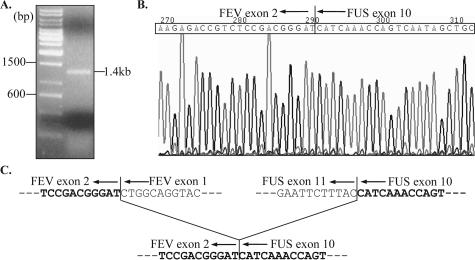
Ewing family tumors are molecularly characterized by expression of chimeric transcripts generated by specific chromosomal translocations, most commonly involving fusion of the EWS gene to a member of the ETS family of transcription factors (including FLI1, ERG, ETV1, E1AF, and FEV). Approximately 85% of reported cases of Ewing sarcoma bear an EWS-FLI1 fusion. In rare cases, FUS can substitute for EWS, with translocation t(16;21)(p11;q24) producing a FUS-ERG fusion with no EWS rearrangement. We report a case of Ewing sarcoma, presenting as a pathological fracture of the distal clavicle in a 33-year-old male, in which cytogenetic analysis revealed a single t(2;16)(q35;p11) balanced translocation. Fluorescence in situ hybridization using a commercially available diagnostic probe was negative for an EWS gene rearrangement; instead, break-apart fluorescence in situ hybridization probes for FUS and FEV were positive for a translocation involving these genes. Cloning and sequencing of the breakpoint region demonstrated an in-frame fusion of FUS to FEV. In conclusion, this represents the first reported case of Ewing family tumors demonstrating a variant translocation involving FUS and FEV and highlights the need to consider alternative permutations of fusion partners for molecular diagnosis of sarcomas.
Figures


Comment in
-
Molecular diagnosis of ewing family tumors: too many fusions... ?J Mol Diagn. 2007 Sep;9(4):437-40. doi: 10.2353/jmoldx.2007.070080. Epub 2007 Jul 25. J Mol Diagn. 2007. PMID: 17652636 Free PMC article.
References
-
- Grier HE. The Ewing family of tumors. Ewing’s sarcoma and primitive neuroectodermal tumors. Pediatr Clin North Am. 1997;44:991–1004. - PubMed
-
- Cheung CC, Kandel RA, Bell RS, Mathews RE, Ghazarian DM. Extraskeletal Ewing sarcoma in a 77-year-old woman. Arch Pathol Lab Med. 2001;125:1358–1360. - PubMed
-
- Folpe AL, Goldblum JR, Rubin BP, Shehata BM, Liu W, Dei Tos AP, Weiss SW. Morphologic and immunophenotypic diversity in Ewing family tumors: a study of 66 genetically confirmed cases. Am J Surg Pathol. 2005;29:1025–1033. - PubMed
-
- O’Sullivan MJ, Perlman EJ, Furman J, Humphrey PA, Dehner LP, Pfeifer JD. Visceral primitive peripheral neuroectodermal tumors: a clinicopathologic and molecular study. Hum Pathol. 2001;32:1109–1115. - PubMed
-
- Khoury JD. Ewing sarcoma family of tumors. Adv Anat Pathol. 2005;12:212–220. - PubMed
Publication types
MeSH terms
Substances
LinkOut - more resources
Full Text Sources
Other Literature Sources
