. 2007 Jul 16;178(2):231-44.
doi: 10.1083/jcb.200611081. Epub 2007 Jul 9.

UXT is a novel and essential cofactor in the NF-kappaB transcriptional enhanceosome

Affiliations

UXT is a novel and essential cofactor in the NF-kappaB transcriptional enhanceosome

Shaogang Sun et al. J Cell Biol. .

Abstract

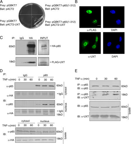
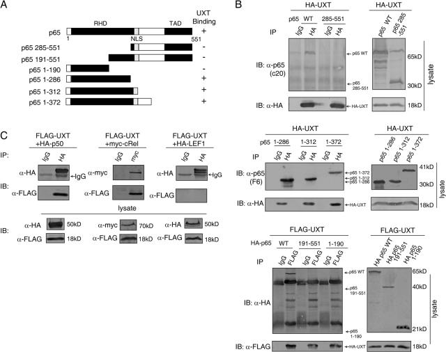
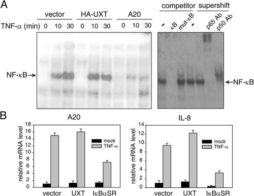
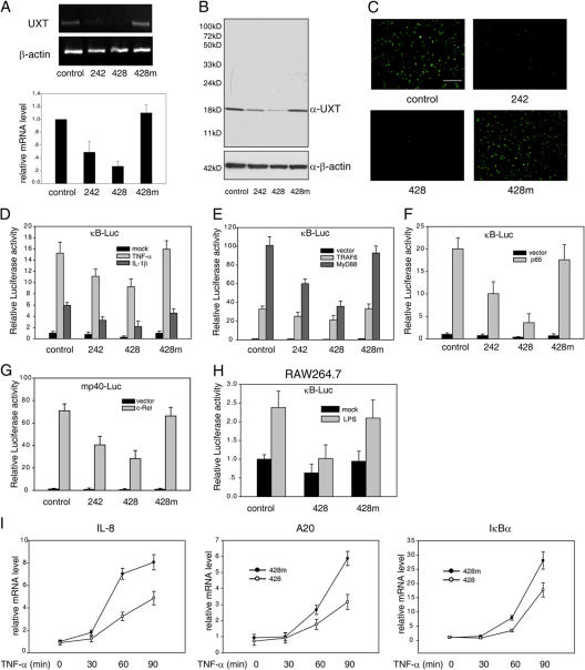
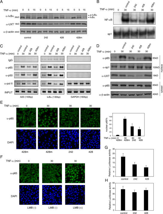
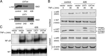
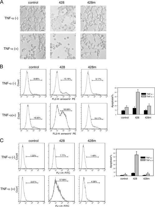
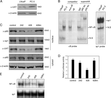
As a latent transcription factor, nuclear factor kappaB (NF-kappaB) translocates from the cytoplasm into the nucleus upon stimulation and mediates the expression of genes that are important in immunity, inflammation, and development. However, little is known about how it is regulated inside the nucleus. By a two-hybrid approach, we identify a prefoldin-like protein, ubiquitously expressed transcript (UXT), that is expressed predominantly and interacts specifically with NF-kappaB inside the nucleus. RNA interference knockdown of UXT leads to impaired NF-kappaB activity and dramatically attenuates the expression of NF-kappaB-dependent genes. This interference also sensitizes cells to apoptosis by tumor necrosis factor-alpha. Furthermore, UXT forms a dynamic complex with NF-kappaB and is recruited to the NF-kappaB enhanceosome upon stimulation. Interestingly, the UXT protein level correlates with constitutive NF-kappaB activity in human prostate cancer cell lines. The presence of NF-kappaB within the nucleus of stimulated or constitutively active cells is considerably diminished with decreased endogenous UXT levels. Our results reveal that UXT is an integral component of the NF-kappaB enhanceosome and is essential for its nuclear function, which uncovers a new mechanism of NF-kappaB regulation.


Figures

Figure 1.
Figure 1.
UXT interacts with p65 in vitro and in vivo. (A) Interaction between p65 and UXT in a yeast two-hybrid assay. (B) Subcellular localization of endogenous and exogenous UXT. 293T cells were transfected with (top) or without (bottom) FLAG-UXT. Immunofluorescentmicroscopy was performed with the indicated primary antibodies. (C) Full-length HA-p65 and FLAG-UXT proteins were labeled with [35S]methionine by in vitro translation. The products were mixed and immunoprecipitated with the indicated antibodies coupled onto protein A/G beads. The immunoprecipitates were resolved by SDS-PAGE and visualized by autoradiography. 10% of the input proteins for pull down are shown at the left. (D) 293T cells were transfected with HA-UXT. 24 h after transfection, cells were treated with 10 ng/ml TNF-α for the indicated times and fractionated to cytoplasmic and nuclear fractions, which were immunoprecipitated and immunoblotted with the indicated antibodies, respectively. (E) 293T cells were treated with 10 ng/ml TNF-α for the indicated times. Whole cell lysates were immunoprecipitated and immunoblotted with the indicated antibodies. Bar, 10 μm.
Figure 2.
Figure 2.
RHD of NF-κB mediates its interaction with UXT. (A) Schematic illustration of p65 and its mutants. RHD, Rel homology domain; NLS, nuclear localization signal; TAD, transactivation domain. (B) Tagged full-length UXT was transfected into 293T cells along with p65 and its deletion mutants as indicated. Whole cell lysates were immunoprecipitated and immunoblotted with the indicated antibodies. (C) 293T cells were transfected with FLAG-UXT together with HA-p50, myc-cRel, and HA–lymphoid enhancer binding factor 1. Cell lysates were immunoprecipitated and immunoblotted with the indicated antibodies.
Figure 3.
Figure 3.
Overexpression of UXT does not markedly affect NF-κB activation induced by TNF-α. (A) 293T cells were transfected with an equal amount of UXT, A20, or control vector. 24 h after transfection, cells were stimulated with 10 ng/ml TNF-α for the indicated times. Equal amounts (10 μg) of nuclear extracts were subjected to EMSA. For competition analysis, 100-fold excess of unlabeled wild-type or mutant κB probes were added to the reaction mixtures. For supershift assays, nuclear extracts were incubated with antibody as indicated. (B) 293T cells were transfected with equal amounts of UXT, IκBαSR, or empty vector. 24 h after transfection, cells were stimulated with 10 ng/ml TNF-α for 30 min or left untreated. Relative mRNA levels of A20 and IL-8 were analyzed by real-time RT-PCR. Data represent means ± SD (error bars) of at least three independent experiments.
Figure 4.
Figure 4.
Knockdown of UXT attenuates NF-κB activation induced by specific stimuli. (A) 293T cells were transfected with the indicated siRNA. 48 h after transfection, the endogenous expression of UXT mRNA was monitored by RT-PCR (top) or by real-time RT-PCR (bottom). (B) Cell lysates from A were subjected to Western blotting for determining endogenous UXT protein levels after siRNA transfection. (C) FLAG-UXT was cotransfected with the indicated siRNA. 24 h after transfection, UXT protein levels were monitored by immunofluorescence. (D) 293T cells were cotransfected with 3×κB-Luc and siRNA as indicated. 48 h after transfection, cells were stimulated with 10 ng/ml TNF-α or 20 ng/ml IL-1 for 7 h before luciferase assays were performed. (E) The indicated siRNA and 3×κB-Luc were transfected into 293T cells along with MyD88 or TRAF6. 48 h after transfection, cells were assayed as in D. (F) p65 was transfected into 293T cells along with the indicated siRNA and 3×κB-Luc. Cells were treated and assayed as in E. (G) c-Rel was transfected into 293T cells along with the indicated siRNA and mp40-Luc. Cells were treated and assayed as in E. (H) RAW264.7 cells were cotransfected with 3×κB-Luc and the indicated siRNA. 48 h after transfection, cells were stimulated with 500 ng/ml lipopolysaccharide for 7 h before luciferase assays were performed. (I) 293T cells were transfected with the indicated siRNA and stimulated by 10 ng/ml TNF-α for the indicated times. Endogenous mRNA expressions of IL-8, A20, and IκBα were measured by real-time RT-PCR. Data represent means ± SD (error bars) of at least three independent experiments. Bar, 200 μm.
Figure 5.
Figure 5.
Knockdown of UXT attenuates the activity and amount of nuclear NF-κB. (A) 293T cells were transfected with the indicated siRNAs. After 48 h, cells were induced by 10 ng/ml TNF-α for the indicated times. Western blotting was performed on the cell extracts to check the phosphorylation and degradation of IκBα. (B) 293T cells were treated as in A. EMSA was performed to test endogenous NF-κB or sp1 binding to their cognate probes. (C) 293T cells were treated as in A. The ChIP assays were performed in terms of A20, IκBα, or GAPDH promoters using antibodies and corresponding primers as described. (D) 293T cells were transfected with the indicated siRNAs. After 48 h, cells were induced by 10 ng/ml TNF-α for 30 min. Cytoplasmic and nuclear fractions were prepared and immunoblotted with the indicated antibodies, respectively. (E) Cells were treated as in D and stained with anti-p65 primary antibody and FITC-conjugated secondary antibody. The nucleus was counterstained with DAPI. Quantification was performed to 100–200 cells in the same ranges of microscopy field for the presence of an appreciable nuclear signal of p65. Only those showing typical focused nuclear p65 were counted. The data are presented as percentages of cells with nuclear p65 versus total cells. (F) 293T cells were transfected with siRNA 428. After 48 h, cells were induced by 10 ng/ml TNF-α or 10 ng/ml TNF-α plus 20 ng/ml LMB for the indicated times and stained with anti-p65 primary antibody and FITC-conjugated secondary antibody. The nucleus was counterstained with DAPI. (G) 293T cells were cotransfected with the indicated siRNAs, 3×κB-Luc, and chimeric p65 (1–312)-VP16. Luciferase assays were performed 48 h after transfection. (H) 293T cells were cotransfected with the indicated siRNAs, Gal4-Luc, and chimeric Gal4 BD-p65 (285–551). Luciferase assays were performed 48 h after transfection. Data represent means ± SD (error bars) of at least three independent experiments. Bars, 20 μm.
Figure 6.
Figure 6.
Rescue of the UXT knockdown effects with a siRNA-resistant form. (A) The HA-tagged wild-type UXT and siRNA-resistant UXT (UXTr) were cotransfected with the indicated siRNAs. After 24 h, cell lysates were subjected to Western blotting for determining exogenous UXT protein levels. (B) 293T cells were transfected with the indicated exogenous UXT and siRNAs. After 48 h, cells were induced by 10 ng/ml TNF-α for the indicated times. Cytoplasmic and nuclear fractions were prepared and immunoblotted with the indicated antibodies, respectively. (C) 293T cells were treated as in B. EMSA was performed to test endogenous NF-κB or sp1 binding to their cognate probes.
Figure 7.
Figure 7.
UXT forms a dynamic complex with NF-κB and is recruited to the NF-κB enhanceosome upon stimulation. (A and B) 293T cells transfected with (A) or without (B) UXT. After 10 ng/ml TNF-α stimulation, ChIP assays were performed on A20, IκBα, or GAPDH promoters as described. (C) 293T cells were induced by 10 ng/ml TNF-α for 30 min, and nuclear extracts were prepared and incubated with the indicated antibodies to perform EMSA supershift assays. (D) 293T cells were transfected with CARM1 siRNA. 48 h after transfection, the endogenous CARM1 mRNA was shown by RT-PCR. (E) 293T cells were transfected with CARM1 or UXT siRNA and treated with 10 ng/ml TNF-α for 30 min or were left untreated. Cover slides were subjected to immunofluoresence assay with anti-p65 antibody. (F) 293T cells were transfected and treated as in E. Nuclear lysates were used for EMSA to check endogenous NF-κB or sp1 DNA binding activities. Bar, 20 μm.
Figure 8.
Figure 8.
Knockdown of UXT sensitizes 293T cells to TNF-α–induced apoptosis. (A) 293T cells were transfected with the indicated siRNAs. 48 h after transfection, cells were treated with 50 ng/ml TNF-α plus 5 μg/ml CHX or were left untreated for 18 h. Representative microscopic images are shown. (B and C) Cells were prepared as in A. After that, cells were analyzed using the annexin V (B) or TUNEL assay (C) to monitor cell apoptosis. The percentages indicate the fractions of positive annexin V cells in total cells. Data represent means ± SD (error bars) of at least three independent experiments. Bar, 10 μm.
Figure 9.
Figure 9.
UXT protein level correlates with the constitutive NF-κB activity in human prostate cancer cell lines. (A) Equal amounts of whole cell lysates from LNcaP or PC-3 prostate cancer cells were immunoblotted with the indicated antibodies. (B) 10 μg of nuclear extracts from LNCaP or PC-3 were subjected to EMSA with a radiolabeled κB or sp1 probe. NS, nonspecific band. (C) PC-3 cells were transfected with the indicated siRNAs. 48 h after transfection, cytoplasmic and nuclear fractions were prepared and immunoblotted with the indicated antibodies, respectively. (D) PC-3 cells were cotransfected with the indicated siRNAs and 3×κB-Luc. Luciferase assays were performed 48 h after transfection. Data represent means ± SD (error) of at least three independent experiments. (E) PC-3 cells were transfected with the indicated siRNAs. 48 h after transfection, nuclear extracts were prepared. Endogenous NF-κB or sp1 DNA binding activities were examined by EMSA.

Similar articles

Cited by

References

    1. Barkett, M., and T.D. Gilmore. 1999. Control of apoptosis by Rel/NF-kappaB transcription factors. Oncogene. 18:6910–6924. - PubMed
    1. Chan, H.M., and N.B. La Thangue. 2001. p300/CBP proteins: HATs for transcriptional bridges and scaffolds. J. Cell Sci. 114:2363–2373. - PubMed
    1. Chen, C.D., and C.L. Sawyers. 2002. NF-kappa B activates prostate-specific antigen expression and is upregulated in androgen-independent prostate cancer. Mol. Cell. Biol. 22:2862–2870. - PMC - PubMed
    1. Chen, F.E., D.B. Huang, Y.Q. Chen, and G. Ghosh. 1998. Crystal structure of p50/p65 heterodimer of transcription factor NF-kappaB bound to DNA. Nature. 391:410–413. - PubMed
    1. Chen, L.F., and W.C. Greene. 2004. Shaping the nuclear action of NF-kappaB. Nat. Rev. Mol. Cell Biol. 5:392–401. - PubMed

Publication types

MeSH terms