. 2007 Jul 16;178(2):201-8.
doi: 10.1083/jcb.200612046. Epub 2007 Jul 9.

The disintegrin and metalloproteinase ADAM12 contributes to TGF-beta signaling through interaction with the type II receptor

Affiliations

The disintegrin and metalloproteinase ADAM12 contributes to TGF-beta signaling through interaction with the type II receptor

Azeddine Atfi et al. J Cell Biol. .

Abstract

Transforming growth factor-beta (TGF-beta) regulates a wide variety of biological processes through two types of Ser/Thr transmembrane receptors: the TGF-beta type I receptor and the TGF-beta type II receptor (TbetaRII). Upon ligand binding, TGF-beta type I receptor activated by TbetaRII propagates signals to Smad proteins, which mediate the activation of TGF-beta target genes. In this study, we identify ADAM12 (a disintegrin and metalloproteinase 12) as a component of the TGF-beta signaling pathway that acts through association with TbetaRII. We found that ADAM12 functions by a mechanism independent of its protease activity to facilitate the activation of TGF-beta signaling, including the phosphorylation of Smad2, association of Smad2 with Smad4, and transcriptional activation. Furthermore, ADAM12 induces the accumulation of TbetaRII in early endosomal vesicles and stabilizes the TbetaRII protein presumably by suppressing the association of TbetaRII with Smad7. These results define ADAM12 as a new partner of TbetaRII that facilitates its trafficking to early endosomes in which activation of the Smad pathway is initiated.


Figures

Figure 1.
Figure 1.
TβRII interacts with ADAM12. (A) Schematic diagram of ADAM12 fragments that interact with TβRII in two-hybrid assays. (B) 293T cells were transfected with HA-TβRII in the presence or absence of Flag-ADAM12. Cell lysates were subjected to anti-Flag immunoprecipitation (IP) followed by immunoblotting (IB) with anti-HA. In this and all of the following experiments, the expression of proteins was determined by direct immunoblotting. (C) C2C12 cells transfected with GFP-ADAM12 and HA-TβRII were immunostained with anti-HA followed by TRITC-conjugated secondary IgG. The panels represent three representative fields. (D–F) Cell extracts from HSC, RD, and C2C12 cells (D) or C2C12 cells (E and F) were immunoprecipitated with anti-ADAM12 (D and F) or anti-TβRII (E) and immunoblotted with the indicated antibodies.
Figure 2.
Figure 2.
ADAM12 increases TGF-β signaling. (A–C) HepG2 (A and B) and C2C12 (C) cells transfected with ARE3-Lux together with FAST1 (A) or CAGA9-Lux (B and C) in the presence or absence of ADAM12 were treated with or without TGF-β. In these and all of the following reporter assays, luciferase activity was determined and normalized, and results are expressed as means ± SD (error bars) of triplicates from five independent experiments. (D) HSC, RD, or C2C12 cells were transfected with the indicated combinations of pEGFP, ADAM12 shRNA, or scrambled shRNA. 36 h later, GFP-transfected cells were sorted by FACS and exposed to TGF-β for 16 h. The expression of endogenous PAI-1, JunB, and ADAM12 was assessed by direct immunoblotting (IB). (E and F) 293T cells were transfected with myc-Smad2, Flag-ADAM12, and HA-Smad4 as indicated. For Smad2 phosphorylation (E), cell lysates were immunoblotted with antiphospho-Smad2. For the association of Smad2 with Smad4 (F), cell lysates were immunoprecipitated (IP) with an anti-myc before immunoblotting with anti-HA. (G) C2C12 cells were transfected with the indicated combinations of pEGFP, ADAM12 shRNA, or scrambled shRNA. 36 h later, GFP-transfected cells were sorted by FACS and exposed to TGF-β for 30 min. The phosphorylation of endogenous Smad2 was assessed by immunoblotting with antiphospho-Smad2. White lines indicate that intervening lanes have been spliced out.
Figure 3.
Figure 3.
Up-regulation of TGF-β signaling by ADAM12 does not involve its cytoplasmic domain or its protease activity. (A–C) HepG2 cells were transfected with CAGA9-Lux, ADAM12, ADAM12-tail, and ADAM12.E351Q as indicated. For A and C, cells were treated with or without TGF-β for 16 h before lysis. Dose effects are shown in insets. For B, cells were treated with 1.10 phenanthroline in the presence or absence of TGF-β. In all cases, luciferase activity was determined. Error bars represent SD. WT, wild type.
Figure 4.
Figure 4.
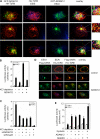
ADAM12 facilitates clathrin-dependent TGF-β signaling. (A) MvLu1 cells transfected with GFP-ADAM12 and HA-TβRII were immunostained with rabbit anti-HA and mouse anti-EEA1 followed by secondary staining with TRITC-conjugated anti–rabbit and Cy5-conjugated anti–mouse IgG. The subcellular localization of GFP-ADAM12 (green), TβRII (red), and EEA1 (blue) was analyzed by a confocal microscope. Colocalization of EEA1 with ADAM12, ADAM12 with TβRII, and EEA1 with TβRII appears as turquoise, yellow, and purple, respectively. The panels represent three independent representative fields. Representative areas from cells that display TβRII and ADAM12 in the EEA1 compartments are enlarged in the insets. (B and C) HepG2 cells were transfected with the indicated combinations of CAGA9-Lux, ADAM12 shRNA, ADAM12, Smad3 shRNA, or scrambled shRNA. Cells were potassium depleted before stimulation with TGF-β, and luciferase activity was examined. (D) COS7 cells were transfected with HA-TβRII and Flag-SARA with or without ADAM12. Cells were immunostained with rabbit anti-HA and mouse anti-EEA1 followed by secondary staining with TRITC-conjugated anti–rabbit and Cy5-conjugated anti–mouse IgG. For the detection of Flag-SARA, cells were incubated with FITC-conjugated anti-Flag. Flag-SARA, TβRII, and EEA1 appear as green, red, and blue, respectively. (E) HepG2 cells transfected with CAGA9-Lux and the indicated expression vectors were treated with nystatin at 10 μM for 1 h before stimulation with TGF-β, and luciferase activity was examined. Error bars represent SD.
Figure 5.
Figure 5.
ADAM12 prevents TβRII degradation. (A) Cells were transfected with HA-TβRII and myc-Ub in the presence or absence of ADAM12. Cell lysates were normalized on the basis of TβRII expression, immunoprecipitated (two samples for each condition) with anti-myc, and immunoblotted with anti-HA. (B) 293T cells were transfected with HA-TβRII and increasing amounts of Flag-ADAM12. Cells were treated with TGF-β for 18 h before lysis, and cell lysates were immunoblotted with anti-HA, anti-Flag, or antiactin. (C) 293T cells transfected with HA-TβRII and the indicated expression vectors were pulse-chased with [35S]Met/Cys, and labeled HA-TβRII was immunoprecipitated and analyzed by SDS-PAGE and autoradiography. (D) Cells were transfected with HA-TβRII, myc-Smad7, and Flag-ADAM12 as indicated. Cell lysates were normalized on the basis of TβRII expression, immunoprecipitated (two samples for each condition) with anti-myc, and immunoblotted with anti-HA. (E) C2C12 cells were transfected with the indicated combinations of pEGFP, ADAM12 shRNA, or scrambled shRNA. 36 h later, GFP-transfected cells were sorted by FACS, lysed, and normalized on the basis of TβRII expression. Then, the association of TβRII with Smad7 or Smad2 was analyzed by immunoprecipitation (IP)/immunoblotting (IB).

Similar articles

Cited by

References

    1. Colland, F., X. Jacq, V. Trouplin, C. Mougin, C. Groizeleau, A. Hamburger, A. Meil, J. Wojcik, P. Legrain, and J.M. Gauthier. 2004. Functional proteomics mapping of a human signaling pathway. Genome Res. 14:1324–1332. - PMC - PubMed
    1. Derynck, R., R.J. Akhurst, and A. Balmain. 2001. TGF-beta signaling in tumor suppression and cancer progression. Nat. Genet. 29:117–129. - PubMed
    1. Di Guglielmo, G.M., C. Le Roy, A.F. Goodfellow, and J.L. Wrana. 2003. Distinct endocytic pathways regulate TGF-beta receptor signalling and turnover. Nat. Cell Biol. 5:410–421. - PubMed
    1. Duffy, M.J., D.J. Lynn, A.T. Lloyd, and C.M. O'Shea. 2003. The ADAMs family of proteins: from basic studies to potential clinical applications. Thromb. Haemost. 89:622–631. - PubMed
    1. Dumont, E., F. Lallemand, C. Prunier, N. Ferrand, A. Guillouzo, B. Clement, A. Atfi, and N. Theret. 2003. Evidence for a role of Smad3 and Smad2 in stabilization of the tumor-derived mutant Smad2.Q407R. J. Biol. Chem. 278:24881–24887. - PubMed

Publication types

MeSH terms