. 2007 Aug;17(8):1170-7.
doi: 10.1101/gr.6101007. Epub 2007 Jul 9.

Nucleosome positioning signals in genomic DNA

Affiliations

Nucleosome positioning signals in genomic DNA

Heather E Peckham et al. Genome Res. 2007 Aug.

Abstract

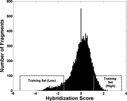
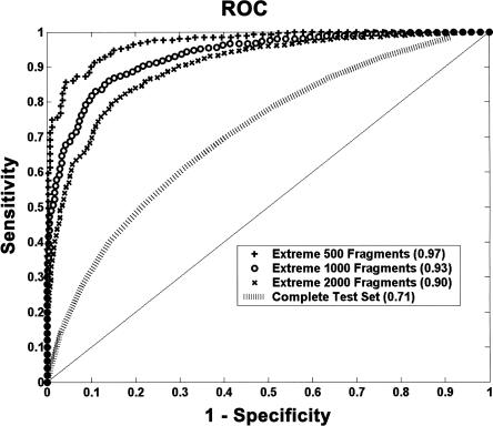
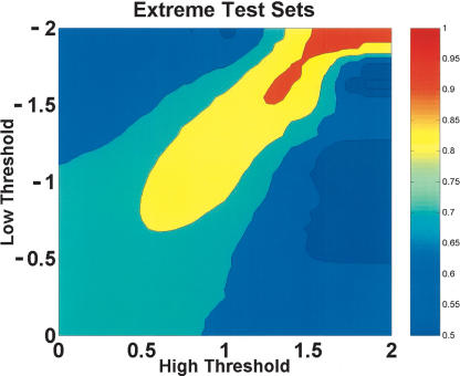
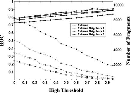
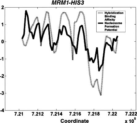
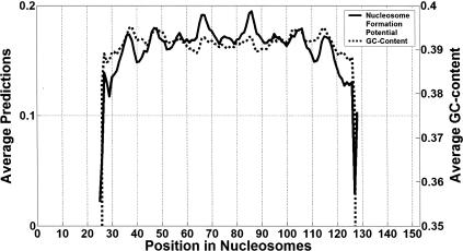
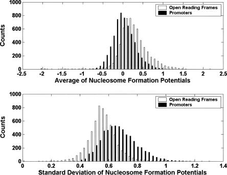
Although histones can form nucleosomes on virtually any genomic sequence, DNA sequences show considerable variability in their binding affinity. We have used DNA sequences of Saccharomyces cerevisiae whose nucleosome binding affinities have been experimentally determined (Yuan et al. 2005) to train a support vector machine to identify the nucleosome formation potential of any given sequence of DNA. The DNA sequences whose nucleosome formation potential are most accurately predicted are those that contain strong nucleosome forming or inhibiting signals and are found within nucleosome length stretches of genomic DNA with continuous nucleosome formation or inhibition signals. We have accurately predicted the experimentally determined nucleosome positions across a well-characterized promoter region of S. cerevisiae and identified strong periodicity within 199 center-aligned mononucleosomes studied recently (Segal et al. 2006) despite there being no periodicity information used to train the support vector machine. Our analysis suggests that only a subset of nucleosomes are likely to be positioned by intrinsic sequence signals. This observation is consistent with the available experimental data and is inconsistent with the proposal of a nucleosome positioning code. Finally, we show that intrinsic nucleosome positioning signals are both more inhibitory and more variable in promoter regions than in open reading frames in S. cerevisiae.


Figures

Figure 1.
Figure 1.
The training set is composed of the fragments with the 1000 highest and the 1000 lowest hybridization scores from chromosome III. High and low hybridization binding affinities indicate that a sequence is nucleosome forming or nucleosome inhibiting, respectively.
Figure 2.
Figure 2.
The ROC scores of the trained support vector machine applied to the test set composed of the fragments not on chromosome III. The support vector machine is trained on the fragments of chromosome III with the most extreme scores and most accurately separates the fragments in the test set with the most extreme scores.
Figure 3.
Figure 3.
The fragments with the strongest nucleosome forming and inhibiting signals are the most accurately predicted within the test set. The X- and Y-axes are the high and low threshold values of the predicted scores of the fragments, respectively. The colors on the contour plot represent the ROC score. For example, at (0, 0) all the fragments in the test set are included and have a ROC score of 0.71, while at (1, −1) only the fragments in the test set with predicted scores ≥1 or ≤−1 are included and have a ROC score of 0.83.
Figure 4.
Figure 4.
The number of fragments in the test set and the corresponding ROC score achieved under the extreme and extreme neighbor conditions. The X-axis represents 0.05 increments in the high threshold, Th. Within each increment of Th, increments of 0.05 from 0.00 to −2.00 of the low threshold, Tl, are computed, and the highest ROC score from these increments is selected. For example, the highest ROC score achieved between a Th of 0 and a Tl between 0.00 and −2.00 is 0.73 from 8264 fragments. The left and right Y-axes represent the ROC score (solid lines) and the number of fragments in the test sets (dashed lines), respectively.
Figure 5.
Figure 5.
The predicted nucleosome formation potentials reproduce the pattern of positioned nucleosomes found with the experimentally determined hybridization binding affinities in the MRM1-HIS3 promoter region.
Figure 6.
Figure 6.
The trained support vector machine reveals the periodicity of the nucleosome formation potential along center-aligned mononucleosomes. The mean predicted nucleosome formation potential over the forward and reverse strands of 199 nucleosome sequences is shown with a solid line, while the GC-content of the sequences is shown with a dashed line. The positions represent the center of the 50-mers on which either the SVM made a prediction or the GC-content was calculated.
Figure 7.
Figure 7.
Intrinsic nucleosome positioning signals are more inhibitory and more variable in promoter regions than in open reading frames in S. cerevisiae. The distributions of the average and standard deviation of the nucleosome formation potentials within each of the promoter regions and open reading frames suggest that promoter regions in S. cerevisiae are specifically designed to inhibit nucleosome formation.

References

    1. Alberts B. Molecular biology of the cell. Garland Science; New York: 2002.
    1. Anselmi C., Bocchinfuso G., De Santis P., Savino M., Scipioni A., Bocchinfuso G., De Santis P., Savino M., Scipioni A., De Santis P., Savino M., Scipioni A., Savino M., Scipioni A., Scipioni A. Dual role of DNA intrinsic curvature and flexibility in determining nucleosome stability. J. Mol. Biol. 1999;286:1293–1301. - PubMed
    1. Baldi P., Brunak S., Chauvin Y., Krogh A., Brunak S., Chauvin Y., Krogh A., Chauvin Y., Krogh A., Krogh A. Naturally occurring nucleosome positioning signals in human exons and introns. J. Mol. Biol. 1996;263:503–510. - PubMed
    1. Boeger H., Griesenbeck J., Strattan J.S., Kornberg R.D., Griesenbeck J., Strattan J.S., Kornberg R.D., Strattan J.S., Kornberg R.D., Kornberg R.D. Nucleosomes unfold completely at a transcriptionally active promoter. Mol. Cell. 2003;11:1587–1598. - PubMed
    1. Boeger H., Griesenbeck J., Strattan J.S., Kornberg R.D., Griesenbeck J., Strattan J.S., Kornberg R.D., Strattan J.S., Kornberg R.D., Kornberg R.D. Removal of promoter nucleosomes by disassembly rather than sliding in vivo. Mol. Cell. 2004;14:667–673. - PubMed

Publication types

LinkOut - more resources