. 2007 Jul 17;104(29):12129-34.
doi: 10.1073/pnas.0701248104. Epub 2007 Jul 9.

Aristolochic acid and the etiology of endemic (Balkan) nephropathy

Affiliations

Aristolochic acid and the etiology of endemic (Balkan) nephropathy

Arthur P Grollman et al. Proc Natl Acad Sci U S A. .

Abstract

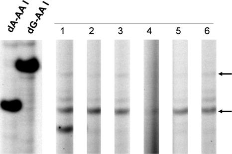
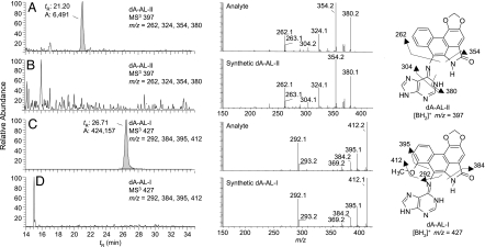
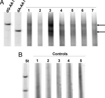
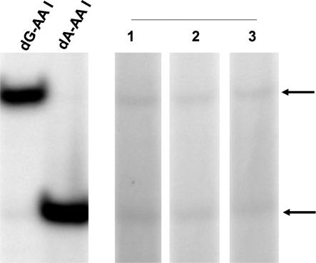
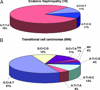
Endemic (Balkan) nephropathy (EN), a devastating renal disease affecting men and women living in rural areas of Bosnia, Bulgaria, Croatia, Romania, and Serbia, is characterized by its insidious onset, invariable progression to chronic renal failure and a strong association with transitional cell (urothelial) carcinoma of the upper urinary tract. Significant epidemiologic features of EN include its focal occurrence in certain villages and a familial, but not inherited, pattern of disease. Our experiments test the hypothesis that chronic dietary poisoning by aristolochic acid is responsible for EN and its associated urothelial cancer. Using (32)P-postlabeling/PAGE and authentic standards, we identified dA-aristolactam (AL) and dG-AL DNA adducts in the renal cortex of patients with EN but not in patients with other chronic renal diseases. In addition, urothelial cancer tissue was obtained from residents of endemic villages with upper urinary tract malignancies. The AmpliChip p53 microarray was then used to sequence exons 2-11 of the p53 gene where we identified 19 base substitutions. Mutations at A:T pairs accounted for 89% of all p53 mutations, with 78% of these being A:T --> T:A transversions. Our experimental results, namely, that (i) DNA adducts derived from aristolochic acid (AA) are present in renal tissues of patients with documented EN, (ii) these adducts can be detected in transitional cell cancers, and (iii) A:T --> T:A transversions dominate the p53 mutational spectrum in the upper urinary tract malignancies found in this population lead to the conclusion that dietary exposure to AA is a significant risk factor for EN and its attendant transitional cell cancer.


Conflict of interest statement

The authors declare no conflict of interest.

Figures

Fig. 1.
Fig. 1.
Detection of AL-DNA adducts in renal tissues of a patient with AAN. DNA (20 μg) was extracted from the renal cortex, medulla, and pelvis of an American woman who developed end-stage renal failure after treatment with an herbal remedy containing Aristolochia. The level of AL-DNA adducts in these tissues was determined by quantitative 32P-postlabeling/PAGE analysis (29). Samples in lanes 1–3 and 4–6 were excised from the right and left kidneys, respectively. Lanes 1 and 4 are from the renal cortex; lanes 2 and 5, from the renal medulla; and lanes 3 and 6, from the renal pelvis. Oligonucleotides containing dA-AL-I and dG-AL-I (1 adduct per 106 dNs), digested in parallel, were used as standards.
Fig. 2.
Fig. 2.
Liquid chromatography electrospray ionization/multistage mass spectrometry (LC-ESI/MS/MSn) analysis of dA-AL adducts in renal tissue. AL-DNA adducts were characterized by LC-ESI/MS/MS3 in the positive ionization mode using a 2-D QIT/MS (LTQ; Thermo Electron, San Jose, CA) interfaced with an Agilent capillary 1100 series LC system (Palo Alto, CA). The MS/MS scan mode was employed to monitor the loss of deoxyribose (dR) from the protonated [M + H − 116]+ to form the protonated base adducts [BH2]+. The consecutive reaction monitoring scan mode in MS3 was used to acquire full product ion spectra of the aglycone ions [BH2]+ of the dA-AL-l and dA-AL-ll adducts. (A and C) DNA from renal cortex of patient described in Fig. 1. (B and D) DNA from renal cortex of a patient who had not been exposed to Aristolochia. DNA (80 μg) was subjected to enzymatic hydrolysis, followed by solid phase extraction enrichment of AL-DNA adducts (47). DNA (20 μg) was injected on column. Mass chromatograms A and B were monitored for dA-AL-II: m/z 513 → 397 → 150–500 Da. C and D were monitored for dA-AL-I: m/z 543: → 427 → 150–500 Da. Chromatograms were reconstructed with the four principal fragment ions observed in the MS3 scan mode. MS3 product ion spectra of dA-AL I and II DNA adducts from human samples and synthetic adduct standards (10 fmol on-column) are illustrated in the middle panel. Proposed mechanisms of fragmentation for each adduct are shown.
Fig. 3.
Fig. 3.
Detection of AL-DNA adducts in the renal cortex. (A) DNA (20 μg) was extracted from seven formalin-fixed, paraffin-embedded renal cortical tissues obtained from four Croatian patients who met the diagnostic criteria for EN and used to quantify AL-DNA adducts by 32P-postlabeling/PAGE techniques. Lanes 1 and 2, patient 1; lane 3, patient 2; lanes 4 and 7, patient 3; lanes 5 and 6, patient 4. (B) Five non-EN patients with upper urothelial cancer; lanes 1–5.
Fig. 4.
Fig. 4.
Identification of AL-DNA adducts in urothelial cancer tissues of patients residing in endemic villages. DNA (10 μg) extracted from upper urinary tract cancer tissues of patients residing in endemic villages of Croatia was used for quantitative analysis of AL-DNA adducts.
Fig. 5.
Fig. 5.
p53 mutational spectra in transitional cell carcinomas. (A) Transitional cell carcinomas from patients with EN (data from Table 1). (B) Transitional cell carcinomas in kidney, renal pelvis, ureter, bladder, and nonspecified urinary organs [data from the IARC p53 database (32).
Fig. 6.
Fig. 6.
Position of p53 base substitution mutations in patients with EN. Arrows above and below the bar indicate mutations observed at G:C and A:T pairs, respectively. Colored arrows represent mutations in the same patient. Single mutations are represented by a black arrow. Numbers corresponding to amino acid residues are shown below the bar.

References

    1. Djukanović L, Radovanović Z. In: Clinical Nephrotoxins. De Broe ME, Porter GA, Bennett WM, Verpooten GA, editors. Dordrecht, The Netherlands: Kluwer Academic; 2003. pp. 587–601.
    1. Stefanović V, Cosyns JP. In: Oxford Textbook of Clinical Nephrology. Davison AM, Cameron JS, Grunfeld JP, Ponticelli C, editors. New York: Oxford Univ Press; 2005. pp. 1095–101.
    1. Tancev I, Evstatijev P, Dorosiev D, Penceva Z, Cvetkov G. Savr Med. 1956;7:14–29. - PubMed
    1. Danilović V. Brit Med J. 1958;1:27–28. - PMC - PubMed
    1. Pichler O, Bobinac E, Miljuš B, Sindik A. Liječ Vjesn. 1959;81:295–306.

Publication types