GPIHBP1: an endothelial cell molecule important for the lipolytic processing of chylomicrons
- PMID: 17620854
- PMCID: PMC2888298
- DOI: 10.1097/MOL.0b013e3281527914
GPIHBP1: an endothelial cell molecule important for the lipolytic processing of chylomicrons
Abstract
Purpose of review: To summarize recent data indicating that glycosylphosphatidylinositol-anchored high density lipoprotein-binding protein 1 (GPIHBP1) plays a key role in the lipolytic processing of chylomicrons.
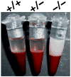
Recent findings: Lipoprotein lipase hydrolyses triglycerides in chylomicrons at the luminal surface of the capillaries in heart, adipose tissue, and skeletal muscle. The endothelial cell molecule that facilitates the lipolytic processing of chylomicrons has never been clearly defined. Mice lacking GPIHBP1 manifest chylomicronemia, with plasma triglyceride levels as high as 5000 mg/dl. In wild-type mice, GPIHBP1 is expressed on the luminal surface of capillaries in heart, adipose tissue, and skeletal muscle. Cells transfected with GPIHBP1 bind both chylomicrons and lipoprotein lipase avidly.
Summary: The chylomicronemia in Gpihbp1-deficient mice, the fact that GPIHBP1 is located within the lumen of capillaries, and the fact that GPIHBP1 binds lipoprotein lipase and chylomicrons suggest that GPIHBP1 is a key platform for the lipolytic processing of triglyceride-rich lipoproteins.
Figures


Similar articles
-
GPIHBP1 and lipolysis: an update.Curr Opin Lipidol. 2009 Jun;20(3):211-6. doi: 10.1097/MOL.0b013e32832ac026. Curr Opin Lipidol. 2009. PMID: 19369870 Free PMC article. Review.
-
Glycosylphosphatidylinositol-anchored high-density lipoprotein-binding protein 1 plays a critical role in the lipolytic processing of chylomicrons.Cell Metab. 2007 Apr;5(4):279-91. doi: 10.1016/j.cmet.2007.02.002. Cell Metab. 2007. PMID: 17403372 Free PMC article.
-
The expression of GPIHBP1, an endothelial cell binding site for lipoprotein lipase and chylomicrons, is induced by peroxisome proliferator-activated receptor-gamma.Mol Endocrinol. 2008 Nov;22(11):2496-504. doi: 10.1210/me.2008-0146. Epub 2008 Sep 11. Mol Endocrinol. 2008. PMID: 18787041 Free PMC article.
-
GPIHBP1, a GPI-anchored protein required for the lipolytic processing of triglyceride-rich lipoproteins.J Lipid Res. 2009 Apr;50 Suppl(Suppl):S57-62. doi: 10.1194/jlr.R800030-JLR200. Epub 2008 Oct 14. J Lipid Res. 2009. PMID: 18854402 Free PMC article. Review.
-
Normal binding of lipoprotein lipase, chylomicrons, and apo-AV to GPIHBP1 containing a G56R amino acid substitution.Biochim Biophys Acta. 2007 Dec;1771(12):1464-8. doi: 10.1016/j.bbalip.2007.10.005. Epub 2007 Oct 22. Biochim Biophys Acta. 2007. PMID: 17997385 Free PMC article.
Cited by
-
Abnormal patterns of lipoprotein lipase release into the plasma in GPIHBP1-deficient mice.J Biol Chem. 2008 Dec 12;283(50):34511-8. doi: 10.1074/jbc.M806067200. Epub 2008 Oct 8. J Biol Chem. 2008. PMID: 18845532 Free PMC article.
-
Syndecan-1 is the primary heparan sulfate proteoglycan mediating hepatic clearance of triglyceride-rich lipoproteins in mice.J Clin Invest. 2009 Nov;119(11):3236-45. doi: 10.1172/JCI38251. Epub 2009 Oct 1. J Clin Invest. 2009. PMID: 19805913 Free PMC article.
-
GPIHBP1 and lipolysis: an update.Curr Opin Lipidol. 2009 Jun;20(3):211-6. doi: 10.1097/MOL.0b013e32832ac026. Curr Opin Lipidol. 2009. PMID: 19369870 Free PMC article. Review.
-
Comparative studies of glycosylphosphatidylinositol-anchored high-density lipoprotein-binding protein 1: evidence for a eutherian mammalian origin for the GPIHBP1 gene from an LY6-like gene.3 Biotech. 2012 Mar;2(1):37-52. doi: 10.1007/s13205-011-0026-4. Epub 2011 Oct 18. 3 Biotech. 2012. PMID: 22582156 Free PMC article.
-
New wrinkles in lipoprotein lipase biology.Curr Opin Lipidol. 2012 Feb;23(1):35-42. doi: 10.1097/MOL.0b013e32834d0b33. Curr Opin Lipidol. 2012. PMID: 22123668 Free PMC article. Review.
References
-
- Havel RJ, Kane JP. Introduction: Structure and metabolism of plasma lipoproteins. In: Scriver CR, Beaudet AL, Sly WS, et al., editors. The Metabolic and Molecular Bases of Inherited Disease. 8. New York: McGraw-Hill; 2001. pp. 2705–2716.
-
- Bengtsson G, Olivecrona T. Activation of lipoprotein lipase by apolipoprotein CII. Demonstration of an effect of the activator on the binding of the enzyme to milk-fat globules. FEBS Lett. 1982;147:183–187. - PubMed
-
- Bengtsson G, Olivecrona T. Lipoprotein lipase: some effects of activator proteins. Eur J Biochem. 1980;106:549–555. - PubMed
-
- Bengtsson G, Olivecrona T. Apolipoprotein CII enhances hydrolysis of monoglycerides by lipoprotein lipase, but the effect is abolished by fatty acids. FEBS Lett. 1979;106:345–348. - PubMed
-
- Kane JP, Havel RJ. Disorders of the biogenesis and secretion of lipoproteins containing the B apolipoproteins. In: Scriver CR, Beaudet AL, Sly WS, et al., editors. The Metabolic and Molecular Bases of Inherited Disease. 8. New York: McGraw-Hill; 2001. pp. 2717–2752.
Publication types
MeSH terms
Substances
Grants and funding
LinkOut - more resources
Full Text Sources
Molecular Biology Databases
Research Materials

