. 2008 May;13(5):498-506.
doi: 10.1038/sj.mp.4002041. Epub 2007 Jul 10.

Glucocorticoid modulation of tryptophan hydroxylase-2 protein in raphe nuclei and 5-hydroxytryptophan concentrations in frontal cortex of C57/Bl6 mice

Affiliations

Glucocorticoid modulation of tryptophan hydroxylase-2 protein in raphe nuclei and 5-hydroxytryptophan concentrations in frontal cortex of C57/Bl6 mice

J A Clark et al. Mol Psychiatry. 2008 May.

Abstract

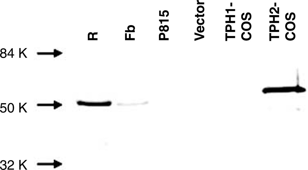
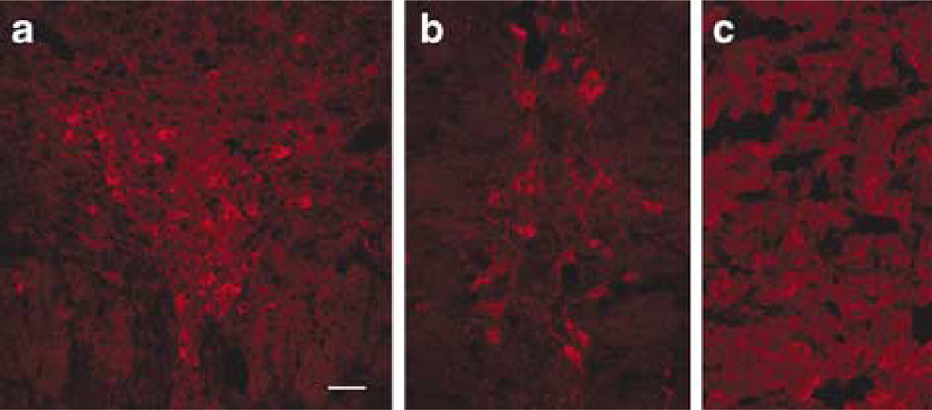
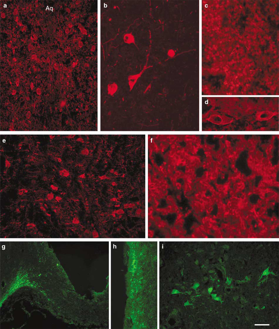
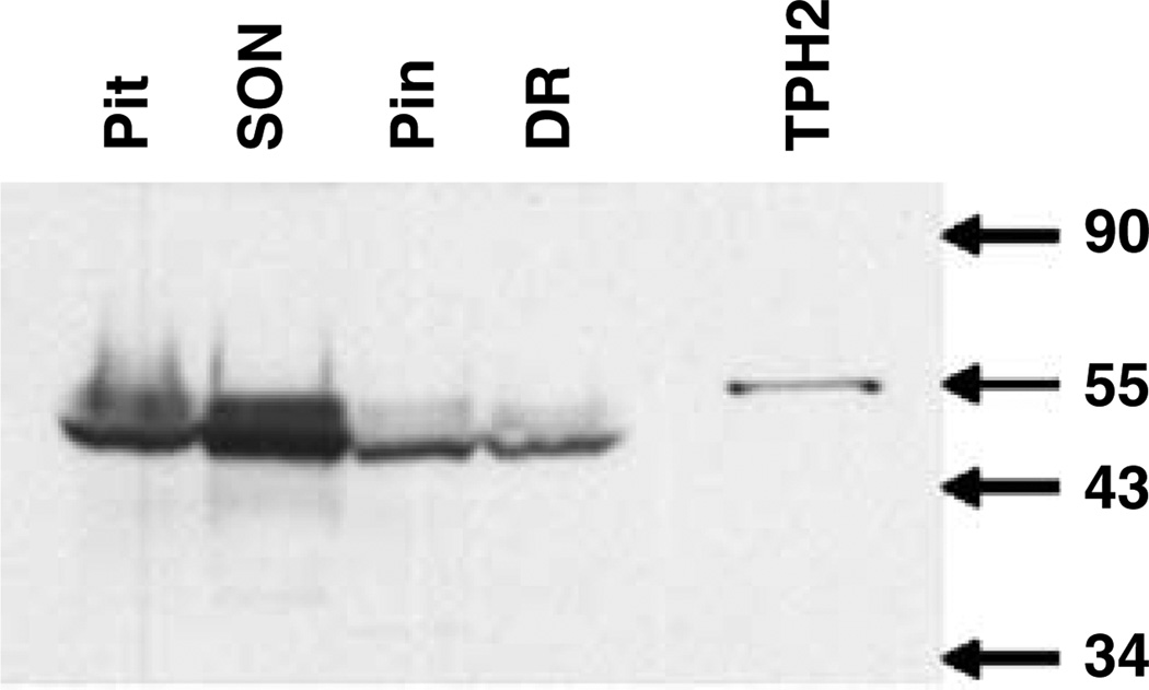
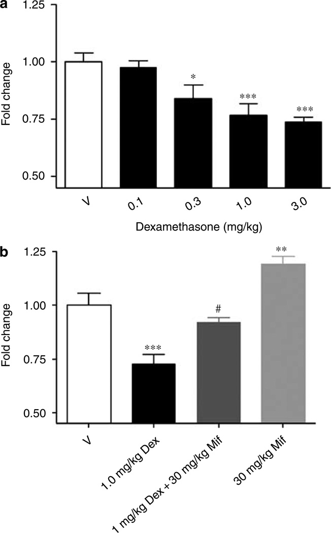
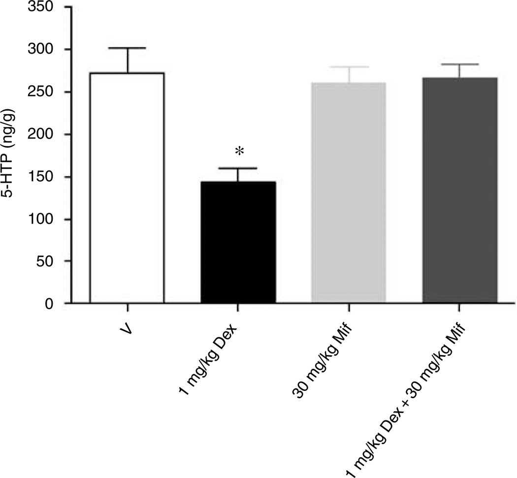
Considerable attention has focused on regulation of central tryptophan hydroxylase (TPH) activity and protein expression. At the time of these earlier studies, it was thought that there was a single central TPH isoform. However, with the recent identification of TPH2, it becomes important to distinguish between regulatory effects on the protein expression and activity of the two isoforms. We have generated a TPH2-specific polyclonal antiserum (TPH2-6361) to study regulation of TPH2 at the protein level and to examine the distribution of TPH2 expression in rodent and human brain. TPH2 immunoreactivity (IR) was detected throughout the raphe nuclei, in lateral hypothalamic nuclei and in the pineal body of rodent and human brain. In addition, a prominent TPH2-IR fiber network was found in the human median eminence. We recently reported that glucocorticoid treatment of C57/Bl6 mice for 4 days markedly decreased TPH2 messenger RNA levels in the raphe nuclei, whereas TPH1 mRNA was unaffected. The glucocorticoid-elicited inhibition of TPH2 gene expression was blocked by co-administration of the glucocorticoid receptor antagonist mifepristone (RU-486). Using TPH2-6361, we have extended these findings to show a dose-dependent decrease in raphe TPH2 protein levels in response to 4 days of treatment with dexamethasone; this effect was blocked by co-administration of mifepristone. Moreover, the glucocorticoid-elicited inhibition of TPH2 was functionally significant: serotonin synthesis was significantly reduced in the frontal cortex of glucocorticoid-treated mice, an effect that was blocked by mifepristone co-administration. This study provides further evidence for the glucocorticoid regulation of serotonin biosynthesis via inhibition of TPH2 expression, and suggest that elevated glucocorticoid levels may be relevant to the etiology of psychiatric diseases, such as depression, where hypothalamic-pituitary-adrenal axis dysregulation has been documented.


Figures

Figure 1
Figure 1
Immunoblotting with TPH2-specific antiserum. Rodent brain tissue samples (40 µg), P815 whole-cell homogenate (30 µg) and COS whole-cell homogenates (1 µg) were run on sodium dodecylsulfate-polyacrylamide gel electrophoresis and transferred to nitrocellulose for immunoblotting. The TPH2-specific antiserum TPH2-6361 detected a dominant ~55 kDa and a minor ~47.5 kDa band in whole-cell homogenates from COS cells overexpressing TPH2 (TPH2-COS). The antiserum detected an ~51 kDa band in total protein from a murine midbrain slice containing raphe nuclei (R) and murine forebrain (Fb). The antiserum did not detect bands in whole-cell homogenates from pcDNA 3.1-transfected COS cells (Vector), COS cells overexpressing TPH1 (TPH1-COS) or in P815 cells, a murine mastocytoma cell line that expresses TPH1 exclusively. TPH, tryptophan hydroxylase.
Figure 2
Figure 2
Immunocytochemical staining of murine central nervous system tissue with TPH2-6361. TPH2-6361 IR was found in neuronal cell bodies in murine dorsal raphe nucleus (a), raphe magnus (b) and pineal gland (c). Arrows point to TPH2 immunoreactivity in the basal artery at the pontine level (b) and in beaded fibers in the pineal body (c) AQ, aqueduct; Bar = 100 µm (a) and 25 µm (b and c). IR, immunoreactivity; TPH, tryptophan hydroxylase.
Figure 3
Figure 3
Immunocytochemical staining of rat central nervous system tissue with TPH2-6361. TPH2-6361 immunoreactivity was found along the raphe nuclei in the midbrain and brainstem. Neuronal cell bodies in rat dorsal raphe nucleus (a), linear raphe (b) and pineal body (c) are shown. Bar = 50 µm. TPH, tryptophan hydroxylase.
Figure 4
Figure 4
Immunocytochemical staining of human central nervous system tissue with TPH2-6361. TPH2-6361 IR in human dorsal raphe neurons at low (a) and high (b) magnification. Panels show TPH2-6361 IR in fibers and cells in the pineal body (c), and pituitary gland (f), blood vessels (d), neurons in the pontine raphe (e) and lateral hypothalamus (i), as well as a fiber network in the median eminence (g) and periventricular zone (h). The fluorochrome used was AlexaFluor-594 (a–f) or AlexaFluor-488 (g–i) Aq, aqueduct. Bar = 25 µm (b), 50 µm (a, e, i) and 100 µm (c, d, fh). IR, immunoreactivity; TPH, tryptophan hydroxylase.
Figure 5
Figure 5
Immunoblotting of tryptophan hydroxylase (TPH)2 in microdissected human brain regions. Total protein from human brain microdissected regions (100 µg) and COS whole-cell homogenate (0.5 µg) were run on sodium dodecylsulfate-polyacrylamide gel electrophoresis and transferred to nitrocellulose for immunoblotting. TPH2-6361 detected a single band of 55 kDa in whole-cell homogenate from COS cells overexpressing TPH2. A predominant band of 47 kDa and a weaker band of 51 kDa was found in total protein from pituitary, supraoptic nucleus, pineal body and dorsal raphe. DR, dorsal raphe; Pin, pineal body; Pit, pituitary; SON supraoptic nucleus; TPH2, COS cell lysate overexpressing TPH2.
Figure 6
Figure 6
Regulation of TPH2 protein by dexamethasone in murine pontine raphe as measured by western blot analyses. Administration (s.c.) of dexamethasone (Dex) at doses from 0.1 to 3.0 mg/kg once daily for 4 days resulted in a reduction in TPH2 protein levels from 23% at 0.3 mg/kg to 30% at 3 mg/kg in the murine raphe slice preparation (a). Administration of 1 mg/kg dexamethasone once daily for 4 days resulted in a 27% decrease in TPH2 protein in the murine raphe slice (b). Co-administration of 1 mg/kg dexamethasone and 30 mg/kg mifepristone (Mif) resulted in protein levels that were not statistically different from vehicle control levels, but were statistically different from the effect of 1 mg/kg dexamethasone alone (b). A 30 mg/kg portion of mifepristone caused a small, but statistically significant increase in TPH2 protein levels (b). *P < 0.01, **P < 0.005, and ***P ≤ 0.001 with respect to vehicle control; #P < 0.005 with respect to 1 mg/kg dexamethasone, one-way analysis of variance. TPH, tryptophan hydroxylase.
Figure 7
Figure 7
In vivo 5-HT synthesis is modulated by dexamethasone in murine frontal cortex. Accumulation of 5-HTP in vivo in murine frontal cortex was reduced by 54% in response to once daily dosing with 1.0 mg/kg dexamethasone for 4 days. Co-administration of mifepristone (30 mg/kg) blocked the effect of dexamethasone (1 mg/kg) on cortical 5-HTP levels and administration of mifepristone alone had no significant effect. *P < 0.01 in comparison to vehicle, one-way analysis of variance + Dunnett’s.

References

    1. Grahame-Smith DG. Tryptophan hydroxylation in brain. Biochem Biophys Res Commun. 1964;16:586–592. - PubMed
    1. Lovenberg W, Jequier E, Sjoerdsma A. Tryptophan hydroxylation: measurement in pineal gland, brainstem, and carcinoid tumor. Science. 1967;155:217–219. - PubMed
    1. Jequier E, Robinson DS, Lovenberg W, Sjoerdsma A. Further studies on tryptophan hydroxylase in rat brainstem and beef pineal. Biochem Pharmacol. 1969;18:1071–1081. - PubMed
    1. Walther DJ, Peter J-U, Bashammakh S, Hortnagl H, Voits M, Fink H, et al. Synthesis of serotonin by a second tryptophan hydroxylase isoform. Science. 2003;299:76. - PubMed
    1. Patel PD, Pontrello C, Burke S. Robust and tissue-specific expression of TPH2 versus TPH1 in rat raphe and pineal gland. Biol Psychiatry. 2004;55:428–433. - PubMed

Publication types

MeSH terms