. 2007 Aug 6;97(3):315-21.
doi: 10.1038/sj.bjc.6603881. Epub 2007 Jul 10.

Characterisation of the new EpCAM-specific antibody HO-3: implications for trifunctional antibody immunotherapy of cancer

Affiliations

Characterisation of the new EpCAM-specific antibody HO-3: implications for trifunctional antibody immunotherapy of cancer

P Ruf et al. Br J Cancer. .

Abstract

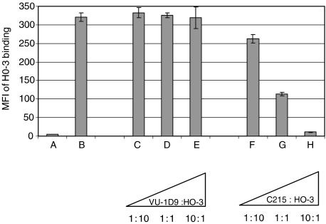
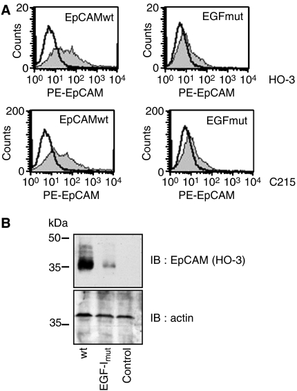
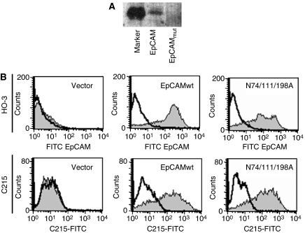
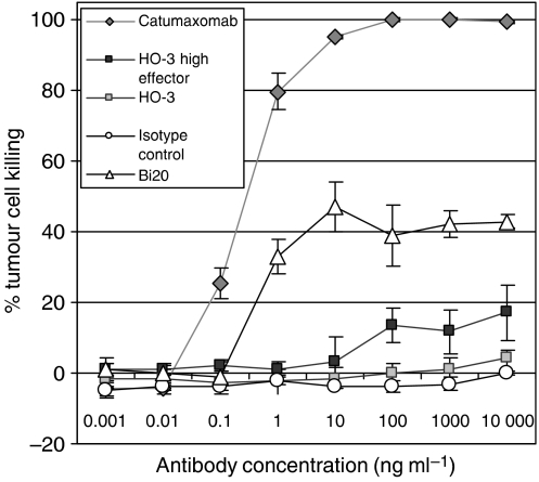
Epithelial cell adhesion molecule EpCAM is a transmembrane glycoprotein that is frequently overexpressed in a variety of carcinomas. This pan-carcinoma antigen has served as the target for a plethora of immunotherapies. Innovative therapeutic approaches include the use of trifunctional antibodies (trAbs) that recruit and activate different types of immune effector cells at the tumour site. The trAb catumaxomab has dual specificity for EpCAM and CD3. In patients with malignant ascites, catumaxomab significantly increased the paracentesis-free interval, corroborating the high efficacy of this therapeutic antibody. Here, we characterised the monoclonal antibody (mAb) HO-3, that is, the EpCAM-binding arm of catumaxomab. Peptide mapping indicated that HO-3 recognises a discontinuous epitope, having three binding sites in the extracellular region of EpCAM. Studies with glycosylation-deficient mutants showed that mAb HO-3 recognised EpCAM independently of its glycosylation status. High-affinity binding was not only detected for mAb HO-3, but also for the monovalent EpCAM-binding arm of catumaxomab with an excellent K(D) of 5.6 x 10(-10) M. Furthermore, trAb catumaxomab was at least a 1000-fold more effective in eliciting the eradication of tumour cells by effector peripheral blood mononuclear cells compared with mAb HO-3. These findings suggest the great therapeutic potential of trAbs and clearly speak in favour of EpCAM-directed cancer immunotherapies.


Figures

Figure 1
Figure 1
C215, but not VU-1D9, competes with mAb HO-3 for binding to wild-type EpCAM. Preincubation of HCT-8 tumour cells with mAb C215 blocked HO-3 binding by more than 50% relative to the positive control (B) with no competitor when the antibodies were added at equal concentrations (2.5 μg ml−1). A 10-fold excess of mAb C215 (25 μg ml−1) resulted in almost complete binding inhibition, which was down to the level of the isotype control (A; IgG2a, kappa). Preincubation of cells with VU-1D9 did not significantly influence HO-3 binding. Triangles indicate escalating amounts of competitor antibody with HO-3 concentration kept constant at 2.5 μg ml−1. Each sample was measured three times; error bars indicate s.d. The experiment was repeated with similar results.
Figure 2
Figure 2
HO-3 binds within EGF-like domain I of EpCAM. (A) HEK293 were transiently transfected with wild-type EpCAM or the EpCAM EGF-I mutant and stained with HO-3 (grey curves; upper panels) or C215 (grey curves; lower panels) in combination with a PE-conjugated secondary antibody. As a control, primary Ab was omitted (black line). (B) Transfections were performed as in (A), proteins were separated by 10% SDS–PAGE, and EpCAM was visualised with the mAb HO-3. For a control, levels of actin were assessed on the same membrane. Shown are the representative results from three independent experiments.
Figure 3
Figure 3
HO-3 and C215 recognise EpCAM independently of the glycosylation status. (A) HEK293 cells were stably transfected with an expression vector for wild-type EpCAM and a triple mutant lacking all glycosylation sites (EpCAMmut). Equal amounts of cell lysates of each cell lines were separated by 10% SDS–PAGE and subjected to glycostaining. Staining was assessed with the FLA 5000 scanning device (Fuji). M represents an internal marker provided by the manufacturers. (B) HEK293 cells stably expressing wild-type EpCAM, a triple mutant lacking all N-glycosylation consensus sites (N74/111/198A; EpCAMmut), or the empty vector for a control were stained with HO-3 (grey curves; upper panels) or C215 (grey curves; lower panels) in combination with a FITC-conjugated secondary antibody. As a control, primary antibody was omitted (black line). All data are representative results from three independent experiments.
Figure 4
Figure 4
TrAb catumaxomab is 1000-fold more potent than HO-3 in eliciting PBMC-mediated killing of EpCAM-positive carcinoma cells. Comparison of trAb and mAb mediated cytotoxicity against EpCAM-positive tumour cells in vitro. HCT-8 tumour cells and PBMCs were co-cultured at ratios of 10 : 1 in the presence of trAbs catumaxomab (anti-EpCAM × anti-CD3), Bi20 (anti-CD20 × anti-CD3) or mAb HO-3 at the indicated concentrations. Additional approaches with high effector ratios of 50 : 1 were performed for H0-3 and mouse IgG2a isotype control MmT1 (anti-mouse Thy-1.2). After 3 days, tumour cell killing was measured by XTT staining. Alloreactivity of PBMC without antibody was not significant (0–2%). Data points display mean values of four determinations with s.d. Data are representative results from four independent experiments using PBMCs from different donors.

Similar articles

Cited by

References

    1. Armstrong A, Eck SL (2003) EpCAM: a new therapeutic target for an old cancer antigen. Cancer Biol Ther 2: 320–326 - PubMed
    1. Baeuerle PA, Gires O (2007) EpCAM (CD326) finding its role in cancer. Br J Cancer 96: 417–423 - PMC - PubMed
    1. Balzar M, Winter MJ, de Boer CJ, Litvinov SV (1999) The biology of the 17-1A antigen (Ep-CAM). J Mol Med 77: 699–712 - PubMed
    1. Bjork P, Jonsson U, Svedberg H, Larsson K, Lind P, Dillner J, Hedlund G, Dohlsten M, Kalland T (1993) - Isolation, partial characterization, and molecular cloning of a human colon adenocarcinoma cell-surface glycoprotein recognized by the C215 mouse monoclonal antibody. J Biol Chem 268: 24232–24241 - PubMed
    1. Chong JM, Speicher DW (2001) Determination of disulfide bond assignments and N-glycosylation sites of the human gastrointestinal carcinoma antigen GA733-2 (CO17-1A, EGP, KS1-4, KSA, and Ep-CAM). J Biol Chem 276: 5804–5813 - PubMed

MeSH terms