. 2007 Jul-Aug;13(7-8):356-61.
doi: 10.2119/2007–00047.Frazzini.

Mild acidosis enhances AMPA receptor-mediated intracellular zinc mobilization in cortical neurons

Affiliations

Mild acidosis enhances AMPA receptor-mediated intracellular zinc mobilization in cortical neurons

Valerio Frazzini et al. Mol Med. 2007 Jul-Aug.

Abstract

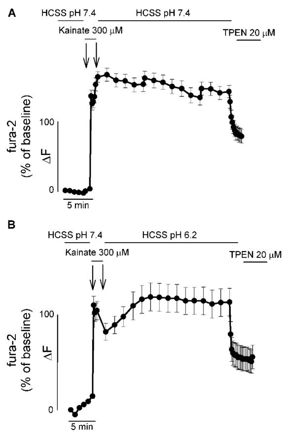
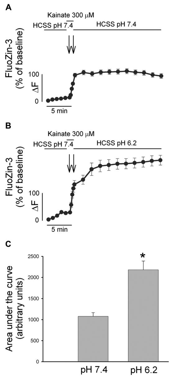
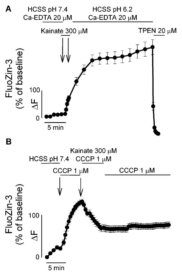
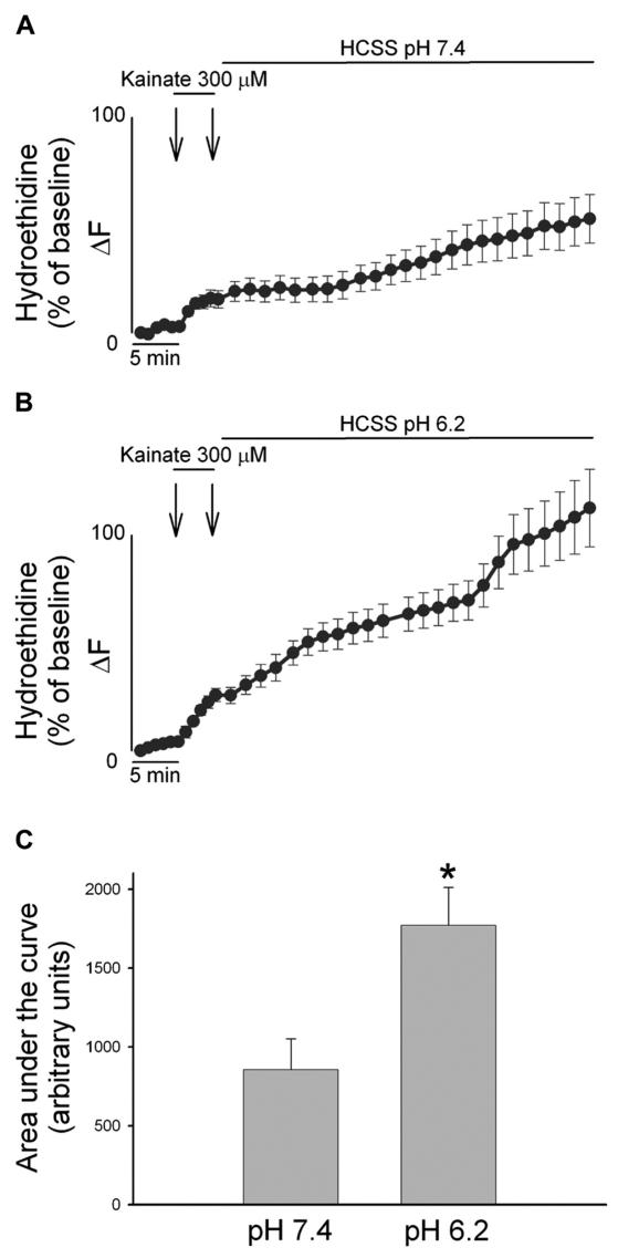
Overactivation of glutamate receptors and subsequent deregulation of the intraneuronal calcium ([Ca2+]i) levels are critical components of the injurious pathways initiated by cerebral ischemia. Another hallmark of stroke is parenchymal acidosis, and we have previously shown that mild acidosis can act as a switch to decrease NMDAR-dependent neuronal loss while potentiating the neuronal loss mediated by AMPARs. Potentiation of AMPAR-mediated neuronal death in an acidotic environment was originally associated only with [Ca2+]i dyshomeostasis, as assessed by Ca2+ imaging; however, intracellular dyshomeostasis of another divalent cation, Zn2+, has recently emerged as another important co-factor in ischemic neuronal injury. Rises in [Zn2+]i greatly contribute to the fluorescent changes of Ca2+-sensitive fluorescent probes, which also have great affinity for Zn2+. We therefore revisited our original findings (Mcdonald et al., 1998) and investigated if AMPAR-mediated fura-2 signals we observed could also be partially due to [Zn2+]i increases. Fura-2 loaded neuronal cultures were exposed to the AMPAR agonist, kainate, in a physiological buffer at pH 7.4 and then washed either at pH 7.4 or pH 6.2. A delayed recovery of fura-2 signals was observed at both pHs. Interestingly this impaired recovery phase was found to be sensitive to chelation of intracellular Zn2+. Experiments with the Zn2+ sensitive (and Ca2+-insensitive) fluorescent probe FluoZin-3 confirmed the idea that AMPAR activation increases [Zn2+]i, a phenomenon that is potentiated by mild acidosis. Additionally, our results show that selective Ca2+ imaging mandates the use of intracellular heavy metal chelators to avoid confounding effects of endogenous metals such as Zn2+.


Figures

Figure 1
Figure 1
AMPAR activation increases both [Ca2+]i and [Zn2+]i levels. A, B. Time course of Ca2+ and Zn2+-dependent changes in fura-2 fluorescence upon activation of AMPARs: Neuronal cultures loaded with fura-2 were imaged before, during, and after a 1 min exposure to 300 μM kainate. After the kainate challenge, neurons were washed for 30 min in a physiological buffer either at pH 7.4 (A) or pH 6.2 (B). At the end of the washout period, neurons were exposed for 5 min to the cell permeant Zn2+ chelator TPEN (20 μM). Traces show mean fura-2 ΔF (expressed as % over baseline of 340/380 nm ratios) (±SEM) of 13–24 neurons from one experiment representative of 5–6, respectively.
Figure 2
Figure 2
Mild acidosis enhances AMPAR-mediated [Zn2+]i rises. A, B. FluoZin-3 loaded cultures were exposed to kainate (300 μM) for 1 min and washed out in a physiological buffer either at pH 7.4 (A) or pH 6.2 (B) for 20 min. Traces show mean FluoZin-3 ΔF (±SEM) of > 50 neurons from 4–7 experiments. C. Bar graph depicts the area under the curve during the recovery phase in the two conditions, * indicates differences between washout at pH 7.4 versus washout at pH 6.2 (P < 0.01). Note that AMPAR-mediated [Zn2+]i rises are greatly enhanced upon exposure to an acidotic environment.
Figure 3
Figure 3
AMPAR-mediated [Zn2+]i rises are mainly dependent on intracellular Zn2+ mobilization. A. AMPAR-mediated [Zn2+]i rises are not modulated by extracellular Zn2+ chelation. FluoZin-3 loaded neurons were exposed, as in Figure 1, to kainate in the presence of the extracellular Zn2+ chelator Ca2+ EDTA (20 μM). Note that the [Zn2+]i rises occurring both during and after kainate exposure are not affected by extracellular chelation of contaminant Zn2+, indicating that the source of these rises is intracellular. B. AMPAR-mediated release of intra-mitochondrial Zn2+. FluoZin-3 loaded cultures were exposed for 5 min to the mitochondrial protonophore, CCCP (1 μM), challenged with 300 μM kainate for 5 min and washed at pH 7.4, in the presence of CCCP. Note that prior exposure to CCCP greatly inhibits subsequent kainate-triggered [Zn2+]i rises, indicating that these two manipulations target a common intracellular Zn2+ pool. Traces show time course of FluoZin-3 Delta;F (±SEM), and are derived from > 15 neurons from 3–5 experiments.
Figure 4
Figure 4
AMPAR-mediated ROS generation is enhanced by low extracellular pH. A, B. Cortical cultures were loaded with HEt, and exposed for 5 min to 300 μM kainate and washed for 50 min with a buffer at pH 7.4 (A) or pH 6.2 (B). HEt fluorescence changes for each neuron are expressed as the ratio of fluorescence at each time point (Fx) to its own baseline fluorescence (F0). Traces show mean (± SEM) of 29 (A) or 40 (B) neurons from 3 experiments. C. Bar graph depicts the cumulative ROS production as area under the curve during the recovery phase in the two conditions, * indicates differences between washout at pH 7.4 versus washout at pH 6.2 (P < 0.01) Note the marked increase in HEt fluorescence only in the case of kainate exposures followed by an acidotic wash out.

Similar articles

Cited by

References

    1. Lee JM, Zipfel GJ, Choi DW. The changing landscape of ischaemic brain injury mechanisms. Nature. 1999;399:A7–14. - PubMed
    1. Chesler M. Regulation and modulation of pH in the brain. Physiol Rev. 2003;83:1183–221. - PubMed
    1. Tang CM, Dichter M, Morad M. Modulation of the N-methyl-D-aspartate channel by extracellular H+ Proc Natl Acad Sci U S A. 1990;87:6445–9. - PMC - PubMed
    1. Traynelis S, Cull-Candy S. Proton inhibition of N-methyl-D-aspartate receptors in cerebellar neurons. Nature. 1990;345:347–50. - PubMed
    1. Giffard RG, Monyer H, Christine CW, Choi DW. Acidosis reduces NMDA receptor activation, glutamate neurotoxicity, and oxygen-glucose deprivation neuronal injury in cortical cultures. Brain Res. 1990;506:339–42. - PubMed

Publication types