

Tuftsin augments antitumor efficacy of liposomized etoposide against fibrosarcoma in Swiss albino mice
- PMID: 17622310
- PMCID: PMC1906688
- DOI: 10.2119/2007–00018.Khan
Tuftsin augments antitumor efficacy of liposomized etoposide against fibrosarcoma in Swiss albino mice
Abstract
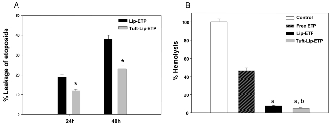
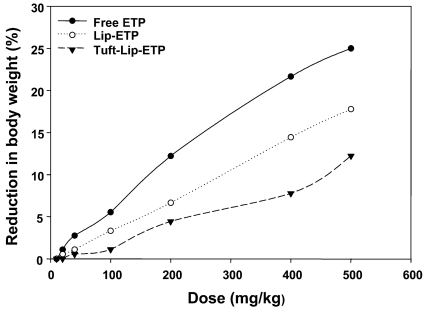
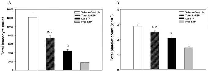
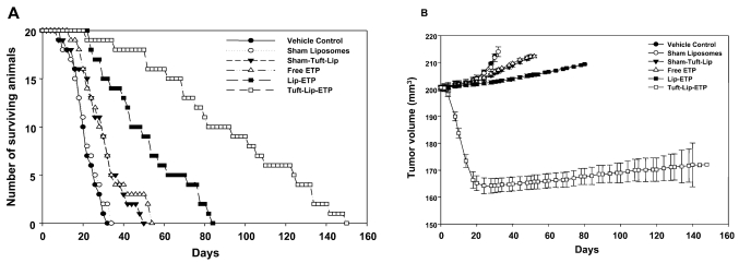
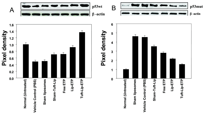
Anticancer drugs are generally plagued by toxic manifestations at doses necessary for control of various forms of cancer. Incorporating such drugs into liposomes not only reduces toxicity but also enhances the therapeutic index. Some antioxidants and potent immunomodulators have also been shown to impart significant antitumor activity presumably by nonspecific activation of the host immune system. In the present study, we evaluated augmentation of the antitumor activity of etoposide (ETP) by the immunomodulator tuftsin in Swiss albino mice with fibrosarcoma. The efficacies of the free form of ETP, liposomized ETP (Lip-ETP), and tuftsin-bearing liposomized ETP (Tuft-Lip-ETP) formulations were evaluated on the basis of tumor regression, effect on expression level of p53wt and p53mut, and survival of the treated animals. Tuft-Lip-ETP, when administered at a dosage of 10 mg/kg body weight/day for five days, significantly reduced tumor volume, delayed tumor growth, and also up-regulated the expression of p53wt. In contrast, although Lip-ETP delayed tumor growth, it did not decrease tumor size. The results of the present study suggest that tuftsin incorporation in drug-loaded liposomes is a promising treatment strategy for various forms of cancers, including fibrosarcoma.
Figures
Similar articles
-
Coadministration of doxorubicin and etoposide loaded in camel milk phospholipids liposomes showed increased antitumor activity in a murine model.Int J Nanomedicine. 2015 Apr 13;10:2847-55. doi: 10.2147/IJN.S80820. eCollection 2015. Int J Nanomedicine. 2015. PMID: 25926730 Free PMC article.
-
Etoposide incorporated into camel milk phospholipids liposomes shows increased activity against fibrosarcoma in a mouse model.Biomed Res Int. 2015;2015:743051. doi: 10.1155/2015/743051. Epub 2015 Mar 2. Biomed Res Int. 2015. PMID: 25821817 Free PMC article.
-
Encapsulation in cationic liposomes enhances antitumour efficacy and reduces the toxicity of etoposide, a topo-isomerase II inhibitor.Pharmacology. 2001;62(3):163-71. doi: 10.1159/000056090. Pharmacology. 2001. PMID: 11287818
-
[Evaluation of antitumor activity of etoposide administered orally for 21 consecutive days against human uterine cancer subcutaneous and/or orthotopic xenografts in nude mice].Gan To Kagaku Ryoho. 1999 Aug;26(9):1313-20. Gan To Kagaku Ryoho. 1999. PMID: 10478185 Japanese.
-
Targeted Drug Delivery Using Tuftsin-bearing Liposomes: Implications in the Treatment of Infectious Diseases and Tumors.Curr Drug Targets. 2021;22(7):770-778. doi: 10.2174/1389450121999201125200756. Curr Drug Targets. 2021. PMID: 33243117 Review.
Cited by
-
Aloe vera induced biomimetic assemblage of nucleobase into nanosized particles.PLoS One. 2012;7(3):e32049. doi: 10.1371/journal.pone.0032049. Epub 2012 Mar 5. PLoS One. 2012. PMID: 22403622 Free PMC article.
-
Immunomodulator effect of picroliv and its potential in treatment against resistant Plasmodium yoelii (MDR) infection in mice.Pharm Res. 2008 Oct;25(10):2312-9. doi: 10.1007/s11095-008-9631-2. Epub 2008 Jun 13. Pharm Res. 2008. PMID: 18551251
-
Mushroom β-Glucan May Immunomodulate the Tumor-Associated Macrophages in the Lewis Lung Carcinoma.Biomed Res Int. 2015;2015:604385. doi: 10.1155/2015/604385. Epub 2015 Jun 17. Biomed Res Int. 2015. PMID: 26167490 Free PMC article.
-
Tuftsin-tailored fusion protein inhibits the growth of circulating gastric tumor cells associated with macrophage phagocytosis.Biochem Biophys Rep. 2023 Feb 24;34:101443. doi: 10.1016/j.bbrep.2023.101443. eCollection 2023 Jul. Biochem Biophys Rep. 2023. PMID: 36875797 Free PMC article.
-
Resveratrol suppresses human cervical carcinoma cell proliferation and elevates apoptosis via the mitochondrial and p53 signaling pathways.Oncol Lett. 2018 Jun;15(6):9845-9851. doi: 10.3892/ol.2018.8571. Epub 2018 Apr 25. Oncol Lett. 2018. PMID: 29928358 Free PMC article.
References
-
- Hande KR. Etoposide: four decades of development of a topoisomerase II inhibitor. Eur J Cancer. 1998;34:1514–21. - PubMed
-
- Spurgers KB, Gold DL, Coombes KR, et al. Identification of cell cycle regulatory genes as principal targets of p53-mediated transcriptional repression. J Biol Chem. 2006;281(35):25134–42. - PubMed
-
- Natalie OK, Tafani M, Rothman RJ, Russo MA, Farber JL. The course of etoposide-induced apoptosis from damage to DNA and p53 activation to mitochondrial release of cytochrome c. J Biol Chem. 2002;277:16547–52. - PubMed
-
- Huang Y, Chan AML, Liu Y, Wang X, Holbrook NJ. Serum withdrawal and etoposide induce apoptosis in human lung carcinoma cell line A549 via distinct pathways. Apoptosis. 1997;2:199–206. - PubMed
Publication types
MeSH terms
Substances
LinkOut - more resources
Full Text Sources