

A structural approach to the assessment of fracture risk in children and adolescents with chronic kidney disease
- PMID: 17622566
- PMCID: PMC6949198
- DOI: 10.1007/s00467-007-0490-6
A structural approach to the assessment of fracture risk in children and adolescents with chronic kidney disease
Abstract
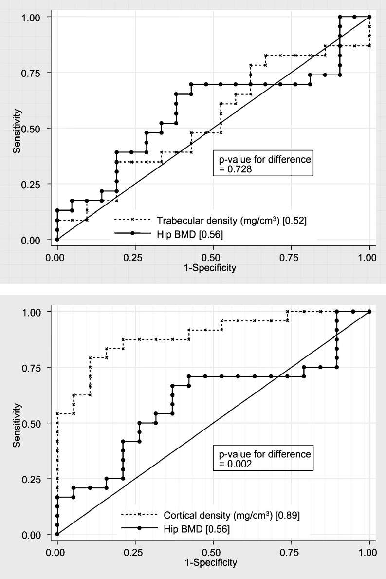
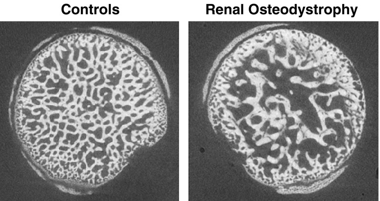
Children with chronic kidney disease (CKD) have multiple risk factors for impaired accretion of trabecular and cortical bone. CKD during childhood poses an immediate fracture risk and compromises adult bone mass, resulting in significantly greater skeletal fragility throughout life. High-turnover disease initially results in thickened trabeculae, with greater bone volume. As disease progresses, resorption cavities dissect trabeculae, connectivity degrades, and bone volume decreases. Increased bone turnover also results in increased cortical porosity and decreased cortical thickness. Dual-energy X-ray absorptiometry (DXA)-based measures of bone mineral density (BMD) are derived from the total bone mass within the projected bone area (g/cm(2)), concealing distinct disease effects in trabecular and cortical bone. In contrast, peripheral quantitative computed tomography (pQCT) estimates volumetric BMD (vBMD, g/cm(3)), distinguishes between cortical and trabecular bone, and provides accurate estimates of cortical dimensions. Recent data have confirmed that pQCT measures of cortical vBMD and thickness provide substantially greater fracture discrimination in adult dialysis patients compared with hip or spine DXA. The following review considers the structural effects of renal osteodystrophy as it relates to fracture risk and the potential advantages and disadvantages of DXA and alternative measures of bone density, geometry, and microarchitecture, such as pQCT, micro-CT (microCT), and micro magnetic resonance imaging (microMRI) for fracture risk assessment.
Figures


References
-
- Parfitt AM. A structural approach to renal bone disease. J Bone Miner Res. 1998;13(8):1213–1220. - PubMed
-
- Alem AM, Sherrard DJ, Gillen DL, Gillen DL, Weiss NS, Beresford SA, Heckbert SR, Wong C, Stelunan-Breen C. Increased risk of hip fracture among patients with end-stage renal disease. Kidney Int. 2000;58(1):396–399. - PubMed
-
- Ball AM, Gillen DL, Sherrard D, Weiss NS, Emerson SS, Seliger SL, Kestenbaum BR, Stelunan-Breen C. Risk of hip fracture among dialysis and renal transplant recipients. JAMA. 2002;288(23):3014–3018. - PubMed
-
- Helenius I, Remes V, Salminen S, Valtra H, Makitie O, Holmberg C, Palmu P, Tervahartiala P, Sarna S, Helnius M, Peltonen J, Jalanko H. Incidence and predictors of fractures in children after solid organ transplantation: a 5-year prospective, population-based study. J Bone Miner Res. 2006;21(3):380–387. - PubMed
-
- Helenius I, Remes V, Tervahartiala P, Salminen S, Sairanen H, Holmberg C, Palmu P, Helenius M, Peltonen J, Jalanko H. Spine after solid organ transplantation in childhood: a clinical, radiographic, and magnetic resonance imaging analysis of 40 patients. Spine. 2006;31(18):2130–2136. - PubMed
Publication types
MeSH terms
LinkOut - more resources
Full Text Sources
Medical
Research Materials
