. 2007 Oct;9(4):424-9.
doi: 10.1215/15228517-2007-023. Epub 2007 Jul 10.

Relationship of glioblastoma multiforme to neural stem cell regions predicts invasive and multifocal tumor phenotype

Affiliations

Relationship of glioblastoma multiforme to neural stem cell regions predicts invasive and multifocal tumor phenotype

Daniel A Lim et al. Neuro Oncol. 2007 Oct.

Abstract

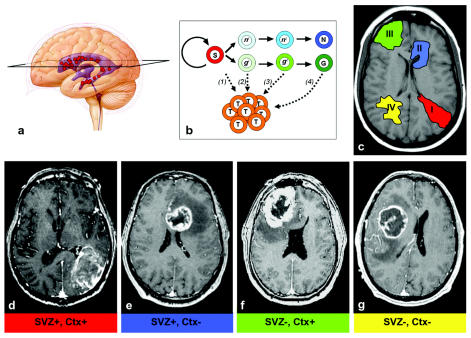
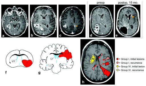
Neural stem cells with astrocyte-like characteristics exist in the human brain subventricular zone (SVZ), and these cells may give rise to glioblastoma multiforme (GBM). We therefore analyzed MRI features of GBMs in specific relation to the SVZ. We reviewed the preoperative and serial postoperative MR images of 53 patients with newly diagnosed GBM. The spatial relationship of the contrast-enhancing lesion (CEL) with the SVZ and cortex was determined preoperatively. Classification was as follows: group I, CEL contacting SVZ and infiltrating cortex; group II, CEL contacting SVZ but not involving cortex; group III, CEL not contacting SVZ but involving cortex; and group IV, CEL neither contacting SVZ nor infiltrating cortex. Patients with group I GBMs (n = 16) were most likely to have multifocal disease at diagnosis (9 patients, 56%, p = 0.001). In contrast, group IV GBMs (n = 14) were never multifocal. Group II (n = 14) and group III (n = 9) GBMs were multifocal in 11% and 29% of cases, respectively. Group I GBMs always had tumor recurrences noncontiguous with the initial lesion(s), while group IV GBM recurrences were always bordering the primary lesion. Group I GBMs may be most related to SVZ stem cells; these tumors were in intimate contact with the SVZ, were most likely to be multifocal at diagnosis, and recurred at great distances to the initial lesion(s). In contrast, group IV GBMs were always solitary lesions; these may arise from non-SVZ, white matter glial progenitors. Our MRI-based classification of GBMs may further our understanding of GBM histogenesis and help predict tumor recurrence pattern.


Figures

Fig. 1
Fig. 1
(A) Neural stem cells (red circles) are found in the walls of the adult brain ventricles (blue). An axial section (black trapezoid) is indicated on this cartoon, representing the approximate level of section of the MR images shown. (B) Multiple hypotheses of brain tumor origin: subventricular zone (SVZ) stem cells (S, red) normally self-renew and produce progenitor cells (light green, glial [g]; light blue, neuronal [n]), which then progress through a developmental lineage (e.g., g1 → g2 …) and differentiate into mature neurons (N, dark blue) or glia (G, dark green). g1 and n1 cells are highly migratory progenitors, directly descendent of SVZ stem cells. g2 and n2 cells represent less migratory local progenitors. Theories include the following: (1) stem cells become dysregulated and give rise to glioma cells (T, orange); (2) highly migratory progenitor cells are the tumor-initiating cells; (3) local progenitors with limited migration give rise to the tumor; and (4) mature glial cells dedifferentiate to produce tumor cells. (C) Schematic of the different groups of glioblastoma multiforme (GBM) lesions based on MRI patterns. (D–G) Axial postcontrast T1-weighted MR images of representative examples for each group (I–IV) in this classification system. (D) For this group I GBM, note that the contrast-enhancing lesion (CEL) extends from the atrium SVZ to the pia. Interestingly, despite the large size, this GBM exerts relatively little mass effect. (E) Group II: SVZ-contacting tumor that does not involve the cortex. (F) Group III: The CEL is invading the cortex, reaching the pia, but does not touch the SVZ. (G) Group IV: These lesions “respect” both the SVZ and cortex; the CEL mass tends to displace brain matter rather than directly invade the cortex and reach the ventricle wall.
Fig. 2
Fig. 2
(A–C) Axial postcontrast T1-weighted MR images of the same patient showing an example of group I multifocal glioblastoma multiforme (GBM). The contrast-enhancing lesion (CEL) contacts the posterior temporal horn subventricular zone (SVZ) in A. Rostrally, there are at least three separate lesions in B and two in C, including one in the contralateral hemisphere (orange asterisk). (D and E) Recurrence of group I GBM in distant SVZ location. (D) Preoperative axial postcontrast T1-weighted MR images of a patient with group I GBM. This patient did not have multifocal disease preoperatively. (E) Fifteen months postoperatively, there was an enhancing mass lesion in the SVZ of the contralateral frontal horn (orange arrowhead) consistent with tumor recurrence. In addition, there is subependymal enhancement bilaterally. No abnormal subarachnoid enhancement was found in this or other images of this patient. (F–H) Integration of GBM cell-of-origin data from animal models with human MRI data. (F) P53, NF1 SVZ astrocytes give rise to group I- and group II-like GBMs in the mouse. This coronal schematic shows an example of a group I tumor (red) extending from the SVZ to the pia. (G) A schematic of human group I and II GBMs in a similar coronal plane. (H) Model of group I and group IV GBM subtypes. At diagnosis, group I GBMs were often multifocal (9 of 16, 56%), possibly from the migration of tumor-initiating cells from either the SVZ or tumor mass. After resection, group I GBM always recurred at locations noncontiguous with the initial lesion(s). Distant, recurrent lesions may arise from tumor cells migrating from the SVZ through the parenchyma (solid arrow) or along the SVZ itself (dotted arrows). The cell of origin of group I GBMs may reside in or near the SVZ, and tumor cells derived from SVZ neural stem cells may have a greater migratory potential than those of group IV. Group IV GBMs occurred only as solitary lesions, and group IV GBM recurrences were always contiguous with the resection cavity. Group IV GBMs may arise from non-SVZ progenitor cells that have a limited migration potential.

Similar articles

Cited by

References

    1. Ignatova TN, Kukekov VG, Laywell ED, Suslov ON, Vrionis FD, Steindler DA. Human cortical glial tumors contain neural stem-like cells expressing astroglial and neuronal markers in vitro. Glia. 2002;39:193–206. - PubMed
    1. Hemmati HD, Nakano I, Lazareff JA, et al. Cancerous stem cells can arise from pediatric brain tumors. Proc Natl Acad Sci U S A. 2003;100:15178–15183. - PMC - PubMed
    1. Galli R, Binda E, Orfanelli U, et al. Isolation and characterization of tumorigenic, stem-like neural precursors from human glioblastoma. Cancer Res. 2004;64:7011–7021. - PubMed
    1. Singh SK, Hawkins C, Clarke ID, et al. Identification of human brain tumour initiating cells. Nature. 2004;432:396–401. - PubMed
    1. Sanai N, Tramontin AD, Quinones-Hinojosa A, et al. Unique astrocyte ribbon in adult human brain contains neural stem cells but lacks chain migration. Nature. 2004;427:740–744. - PubMed