Review
. 2007 Aug;211(2):199-211.
doi: 10.1111/j.1469-7580.2007.00775.x. Epub 2007 Jul 9.

A multiarchitectonic approach for the definition of functionally distinct areas and domains in the monkey frontal lobe

Affiliations
Review

A multiarchitectonic approach for the definition of functionally distinct areas and domains in the monkey frontal lobe

Abdelouahed Belmalih et al. J Anat. 2007 Aug.

Abstract

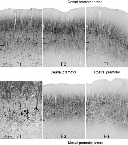
Over the last century, anatomical studies have shown that the cerebral cortex can be subdivided into structurally distinct regions, giving rise to a new branch of neuroanatomy: 'architectonics'. Since then, architectonics has been often accused of being overly subjective, and its validity for the definition of functionally different cortical fields has been seriously questioned. Since the late 1980s, however, the problem of localization has become particularly important in functional studies of the primate motor cortex, because of evidence that (1) the primate motor cortex is made up of a mosaic of functionally specialized areas and (2) the human motor cortex shares several general organizational principles with the monkey motor cortex. Studies of the macaque agranular frontal cortex that used a multimodal cyto-, myelo- and immuno-architectonic approach have shown that architectonic borders can be reliably and consistently defined across different individuals, even at a qualitative level of analysis. The validity of this approach has been confirmed by its ability to localize functionally distinct areas precisely and to predict the existence of new functional areas. After more than a century, architectonics as a discipline goes far beyond its original aim of generating cortical maps.


Figures

Fig. 1
Fig. 1
Low-power photomicrographs of representative fields trough the macaque motor cortex from a parasagittal (A) and a coronal (B,C) section stained via cytocrome oxidase histochemistry. Arrows mark the borders between architectonic areas. The dashed box within the section drawings indicates the location of the photomicrograph. The levels at which the sections were taken are shown in the drawings of dorsal and dorsolateral views of the hemisphere. Scale bar, shown in A, applies also to B and C. (D) Mesial and lateral views of the macaque brain showing the histochemical map of the agranular frontal cortex, according to Matelli et al. (1985). The cingulate sulcus is shown unfolded. AFC, agranular frontal cortex; AI, inferior arcuate sulcus; AS, superior arcuate sulcus; C, central sulcus; Ca, calcarine fissure; Cg, cingulate sulcus; IO, inferior occipital sulcus; IP, intraparietal sulcus; L, lateral fissure; Lu, lunate sulcus; P, principal sulcus; POM, mesial parieto-occipital sulcus; Skc, somatic koniocortex; ST, superior temporal sulcus.
Fig. 2
Fig. 2
Low-power photomicrographs from Nissl-stained parasagittal sections, taken from two different macaque brains, centred on the precentral gyrus. Scale bar, shown in A, applies also to B. Conventions and abbreviations as in Fig. 1.
Fig. 3
Fig. 3
Low-power photomicrographs and higher magnification views of representative fields of cytoarchitectonic areas F1, F2, F3, F6 and F7. Scale bars, shown in the low-power and in the higher magnification view of F1, apply to all low-power and higher magnification views, respectively.
Fig. 4
Fig. 4
Mesial and lateral views of the macaque brain showing the architectonic parcellation of the agranular frontal cortex according to Matelli et al. (1985, 1991). The cingulate sulcus is shown unfolded. On the basis of the available data, the various body-part representations are reported (see, for example, Rizzolatti et al. 1998). In the prefrontal cortex the frontal eye field (FEF) is defined according to physiological criteria (Bruce et al. 1985). F1 (primary motor cortex), caudal (F3, F2, F4 and F5) and rostral (F6 and F7) premotor areas are shown in green, in orange and in blue, respectively. Abbreviations as in Fig. 1.
Fig. 5
Fig. 5
Photomicrographs of representative fields of areas F1, F2, F3, F6 and F7 showing the laminar distribution pattern of SMI-32 immunoreactivity. Scale bar, shown in F1 (upper left), applies to all photomicrographs, except the higher magnification view of F1 (lower left). Borders between layers are reported from an adjacent Nissl-stained section.
Fig. 6
Fig. 6
Low-power photomicrographs of SMI-32-immunostained parasagittal sections through the transition between the agranular with the granular frontal cortex (A) and the superior arcuate sulcus (B). Dashed box within the section drawings indicates the location of the photomicrograph. The levels at which the sections were taken are shown in the drawing of a dorsal view of the hemisphere. Scale bar, shown in A, applies also to B. Abbreviations as in Fig. 1.
Fig. 7
Fig. 7
Upper part: low-power photomicrograph of a CB-immunostained parasagittal section through the whole extent of the motor cortex, taken at a mediolateral level similar to that of Fig. 1(A). Lower part: photomicrographs of representative fields of areas F1, F2, F3, F6 and F7, showing the laminar distribution pattern of CB immunoreactivity. Scale bar, shown in F6, applies to all photomicrographs. Borders between layers are reported from an adjacent Nissl-stained section.
Fig. 8
Fig. 8
Upper part: low-power photomicrograph of an SMI-32-immunostained parasagittal section centred on the precentral gyrus and the whole extent of the inferior arcuate sulcus. The level at which the section was taken is shown in the drawing of a dorsal view of the hemisphere. Lower part: higher magnification views of representative fields of F5p (upper row) and F5a (lower row) showing the cytoarchitecture (right) and the laminar distribution pattern of SMI-32 (middle) and CB (right) immunoreactivity. Borders between layers are reported from an adjacent Nissl-stained section. Scale bar, shown in the upper left photomicrographs, applies to all fields.

References

    1. Baleydier C, Achache P, Froment JC. Neurofilament architecture of superior and mesial premotor cortex in the human brain. Neuroreport. 1997;8:1691–1696. - PubMed
    1. von Bonin G, Bailey P. The Neocortex of Macaca Mulatta. Urbana, IL: University of Illinois Press; 1947.
    1. von Bonin G. Architecture of the precentral motor cortex and some adjacent areas. In: Bucy PC, editor. The Precentral Motor Cortex. Urbana, IL: University of Illinois Press; 1949. pp. 7–82.
    1. Brodmann K. Vergleichende Lokalisationslehre der Groshirnrinde. Leipzig: Barth; 1909.
    1. Bruce CJ, Goldberg ME, Bushnell C, Stanton GB. Primate frontal eye fields. II. Physiological and anatomical correlates of electrically evoked movements. J Neurophysiol. 1985;54:714–734. - PubMed

Publication types

LinkOut - more resources