Comparative Study
. 2007 Jul 10:4:72.
doi: 10.1186/1743-422X-4-72.

Structural comparisons of the nucleoprotein from three negative strand RNA virus families

Affiliations
Comparative Study

Structural comparisons of the nucleoprotein from three negative strand RNA virus families

Ming Luo et al. Virol J. .

Abstract

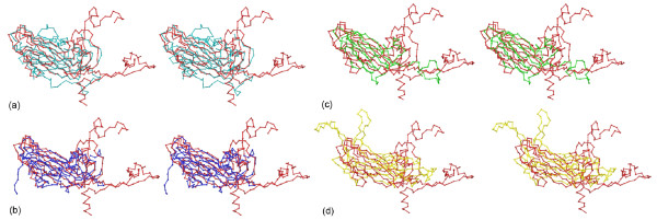
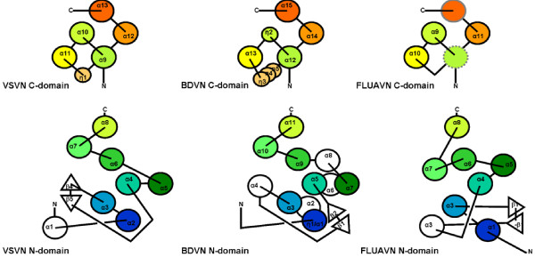
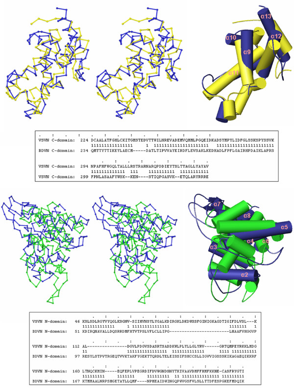
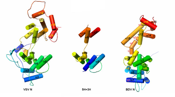
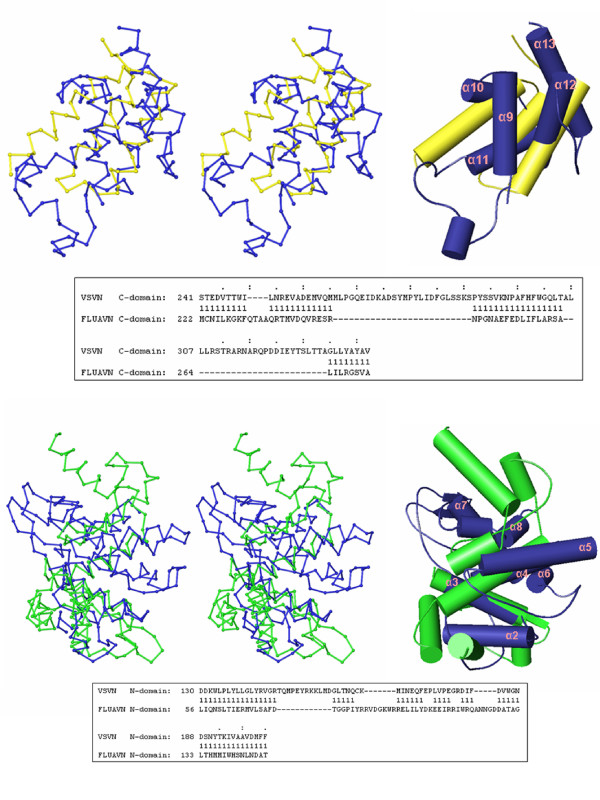
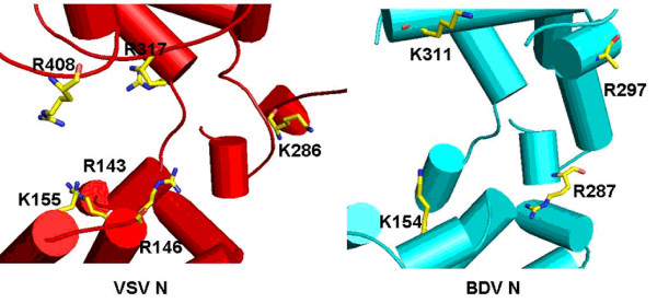
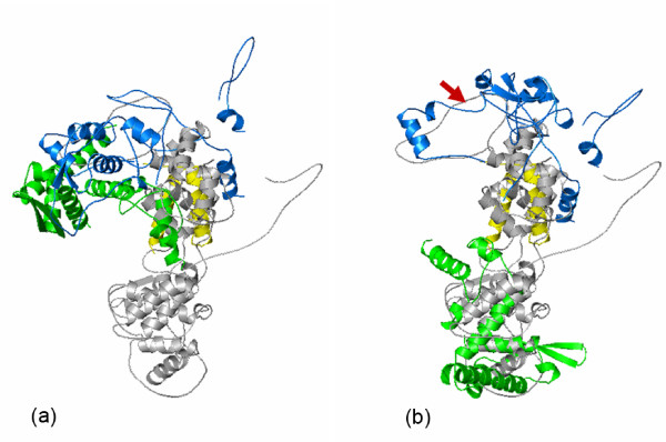
Structures of the nucleoprotein of three negative strand RNA virus families, borna disease virus, rhabdovirus and influenza A virus, are now available. Structural comparisons showed that the topology of the RNA binding region from the three proteins is very similar. The RNA was shown to fit into a cavity formed by the two distinct domains of the RNA binding region in the rhabdovirus nucleoprotein. Two helices connecting the two domains characterize the center of the cavity. The nucleoproteins contain at least 5 conserved helices in the N-terminal domain and 3 conserved helices in the C-terminal domain. Since all negative strand RNA viruses are required to have the ribonucleoprotein complex as their active genomic templates, it is perceivable that the (5H+3H) structure is a common motif in the nucleoprotein of negative strand RNA viruses.


Figures

Figure 1
Figure 1
β-barrel comparisons. (a) Stereo Cα drawings for the superposition of the β-barrel fold in HRV16-VP2 (cyan) with that in HRV16-VP1 (red). (b) SBMV (blue) with HRV16-VP1 (red). (c) STMV (green) with HRV16-VP1 (red). (d) HPV (yellow) with HRV16-VP1 (red). In this and the following figures, the Cα tracing was prepared with RIBBONS [14] and protein structural cartoons were prepared with PyMol [15].
Figure 2
Figure 2
Topology drawings for the C-terminal domain (top panel) and the N-terminal domain of the N proteins. Large circles represent α-helices and triangles represent β-strands. Small circles represent 310 helices. Color codes are from blue to red orange, corresponding to the sequence distance to the N-terminus similar as in Figure 4. Lines above the circles represent connections on top of the helices, whereas lines below the circles represent the connections at bottom of the helices. The secondary structure elements are labeled the same as in the reported crystal structures. The dotted gray colored circle in the FLUAN C-terminal domain implies a possible disordered α-helix and the gray circle implies a mismatch of a loop with an α-helix.
Figure 3
Figure 3
Comparisons of the VSV and BDV N structures. Stereo Cα drawings for the superposition of the C-terminal domain in the VSV N protein (blue) with that of the BDV N protein (yellow) (upper panel), and the N-terminal domain of the VSV N protein (blue) with that of the BDV N protein (green) (lower panel). Residue positions of the aligned structures are shown in the box below each structural comparison. '1' marks the aligned residues between the two structures. Cartoon drawings are also presented on the right with α-helices in VSV N labeled.
Figure 4
Figure 4
The structures of the VSV and the BDV N proteins compared to the (5H+3H) motif. The aligned helices are labeled by black lettering in each structure.
Figure 5
Figure 5
Comparisons of the VSV and FLUAV N structures. Stereo cartoon drawings for the superposition of the C-terminal domain of the FLUAV N protein (yellow) with that of the VSV N protein (blue), and the superposition of the N-terminal domain of the FLUAV N protein (dark green) with that of the VSV N protein (blue). The two domains in the FLUAV N protein were aligned with their counterparts in the VSV N proteins as separate domains. In the case of the N-terminal domain, only the core residues 56–147 were included in the calculation for structural alignment by FATCAT. Residue positions of the aligned structures are shown in the box below each structural comparison. '1' marks the aligned residues between the two structures. Cartoon drawings are also presented on the right with α-helices in VSV N labeled. Statistics for the domain alignments could be found in Table 1B and 1C.
Figure 6
Figure 6
The RNA binding cavity of the VSV N protein (red) with highlighting the positively charged residues that interact with the RNA. For comparison, the similar region in the BDV N protein (cyan) was presented. Positively charged residues that could potentially interact with the RNA are also highlighted in the BDV N structure. The sidechain of Arg297 in the BDV N structure was disordered in the crystal structure (not shown in this figure).
Figure 7
Figure 7
Cartoon drawings for the superposition of the FLUAV N protein with that of the VSV N protein (gray). (a) Superposition of the FLUAV N protein as in the reported crystal structure. Only the C-terminal domain of the putative RNA binding region of the FLUAV N protein was included in the calculation for the structural alignment by FATCAT [11]. The N-terminal domain of the putative RNA binding region in the FLUAV N protein was colored green, the C-terminal domain, yellow, and the addition domain at the C-terminal end of the FLUAV N protein, blue. (b) Superposition of a hypothetical structure of the FLUAV N protein with the structure of the VSV N protein. The orientation of the C-terminal domain is the same in (a) and (b). The N-terminal domain (green) was aligned with the N-terminal domain of the VSV N protein as in Figure 5. The additional domain at the C-terminal end (blue) was positioned by twisting the torsion angles of the peptide chain including residues 295–297 (indicated by the red arrow) to match the loop at the end of the VSV C-terminal domain.

References

    1. Rossmann MG, Johnson JE. Icosahedral RNA virus structur. Annu Rev Biochem. 1989;58:533–573. doi: 10.1146/annurev.bi.58.070189.002533. - DOI - PubMed
    1. Bamford DH, Grimes JM, Stuart DI. What does structure tell us about virus evolution? Curr Opin Struct Biol. 2005;15:655–663. doi: 10.1016/j.sbi.2005.10.012. - DOI - PubMed
    1. Rudolph MG, Kraus I, Dickmanns A, Eickmann M, Garten W, Ficner R. Crystal structure of the borna disease virus nucleoprotein. Structure. 2003;11:1219–1226. doi: 10.1016/j.str.2003.08.011. - DOI - PubMed
    1. Green TJ, Zhang X, Wertz GW, Luo M. Structure of the vesicular stomatitis virus nucleoprotein-RNA complex. Science. 2006;313:357–360. doi: 10.1126/science.1126953. - DOI - PubMed
    1. Albertini AA, Wernimont AK, Muziol T, Ravelli RB, Clapier CR, Schoehn G, Weissenhorn W, Ruigrok RW. Crystal structure of the rabies virus nucleoprotein-RNA complex. Science. 2006;313:360–363. doi: 10.1126/science.1125280. - DOI - PubMed

Publication types

MeSH terms

LinkOut - more resources