. 2007 Jul 10:6:8.
doi: 10.1186/1477-5751-6-8.

Peripheral nervous system manifestations in a Sandhoff disease mouse model: nerve conduction, myelin structure, lipid analysis

Affiliations

Peripheral nervous system manifestations in a Sandhoff disease mouse model: nerve conduction, myelin structure, lipid analysis

Melanie A McNally et al. J Negat Results Biomed. .

Abstract

Background: Sandhoff disease is an inherited lysosomal storage disease caused by a mutation in the gene for the beta-subunit (Hexb gene) of beta-hexosaminidase A (alphabeta) and B (beta beta). The beta-subunit together with the GM2 activator protein catabolize ganglioside GM2. This enzyme deficiency results in GM2 accumulation primarily in the central nervous system. To investigate how abnormal GM2 catabolism affects the peripheral nervous system in a mouse model of Sandhoff disease (Hexb-/-), we examined the electrophysiology of dissected sciatic nerves, structure of central and peripheral myelin, and lipid composition of the peripheral nervous system.

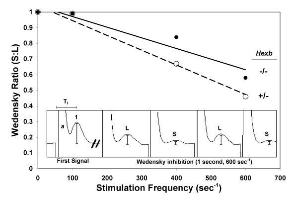
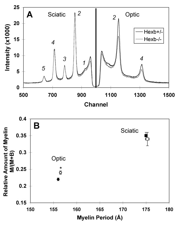
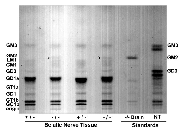
Results: We detected no significant difference in signal impulse conduction velocity or any consistent change in the frequency-dependent conduction slowing and failure between freshly dissected sciatic nerves from the Hexb+/- and Hexb-/- mice. The low-angle x-ray diffraction patterns from freshly dissected sciatic and optic nerves of Hexb+/- and Hexb-/- mice showed normal myelin periods; however, Hexb-/- mice displayed a approximately 10% decrease in the relative amount of compact optic nerve myelin, which is consistent with the previously established reduction in myelin-enriched lipids (cerebrosides and sulfatides) in brains of Hexb-/- mice. Finally, analysis of lipid composition revealed that GM2 content was present in the sciatic nerve of the Hexb-/- mice (undetectable in Hexb+/-).

Conclusion: Our findings demonstrate the absence of significant functional, structural, or compositional abnormalities in the peripheral nervous system of the murine model for Sandhoff disease, but do show the potential value of integrating multiple techniques to evaluate myelin structure and function in nervous system disorders.


Figures

Figure 1
Figure 1
Wedensky Ratio vs. Stimulation Frequency in Hexb+/- and Hexb-/- Mice. Wedensky ratios (see Materials and Methods) for Hexb+/- (○, dashed line) and Hexb-/- (●, solid line) mice were plotted against the stimulation frequency with linear regressions (n = 6–10) to analyze frequency-dependent conduction failure in the two mouse models. As evidenced by the decrease in the Wedensky ratio in both groups, conduction failure after a one second stimulus train increased in alternating CAP signals with increasing stimulation frequency. The slopes of the linear regressions were not different within 95% confidence levels indicating similar conduction failure behavior in the Hexb+/- and Hexb-/- mice. The first CAP signal recorded during a 1 second supramaximal stimulation at 600 sec-1 is compared to the last four CAP signals in the train (scale conserved). Wedensky inhibition is observed. Tl, latency used for CNCV calculations; a, stimulus artifact; 1, amplitude of first CAP in stimulus train; L, S, CAP amplitudes after 1 sec of 600 Hz stimulation (1.67 msec between stimuli).
Figure 2
Figure 2
Diffraction from Optic and Sciatic Nerves in Hexb+/- and Hexb-/- Mice. (A) Representative examples of data for sciatic (left) and optic (right) nerves from Hexb+/- (black) and Hexb-/- (grey) mice. Whereas indistinguishable patterns were obtained for sciatic nerve samples from both groups, optic nerves from Hexb-/- mice showed weaker myelin scatter compared to those from Hexb+/- mice. The Bragg orders for the x-ray peaks are indicated as 1–5. (B) The fraction of total x-ray scatter (M+B) that is accounted for by compact myelin (M) (i.e., M/(M+B)), was plotted against the myelin period (d) [16]. For optic nerve myelin, the Hexb+/- (○) and Hexb-/- (●) mice have similar periods; however, the Hexb-/- mice have less relative myelin in the CNS when compared to the Hexb+/- mice (n = 3–4 per group, p < 0.05; two-tailed, unpaired t-test). For sciatic nerve, the Hexb+/- (□) and Hexb-/- (■) mice have similar periods and relative amounts of compact myelin (n = 8 per group). Thus, x-ray diffraction revealed no myelin abnormalities in the PNS and less relative amounts of compact myelin in the CNS of the Hexb-/- mice.
Figure 3
Figure 3
Myelin Membrane Packing in Optic and Sciatic Nerves from Hexb+/- and Hexb-/- Mice. The integral widths w2 are plotted as a function of h4 to determine the relative amount of myelin packing disorder according to the theory of paracrystalline diffraction [18]. The projected intercept on the ordinate axis is inversely related to the number of repeating units N (the coherent domain size), and the slope is proportional to the fluctuation in period, Δ (lattice or stacking disorder). There were no differences within 95% confidence levels between the Hexb+/- (open symbols, dashed line) and Hexb-/- (filled symbols, solid line) slopes of the optic (circles) or sciatic (squares) nerves (n = 3–8) indicating no change in the membrane packing of the internodal compact myelin for the sciatic nerves (PNS) and for the optic nerves (CNS).
Figure 4
Figure 4
HPTLC of Ganglioside Distribution in Hexb+/- and Hexb-/- Mice. HPTLC of two Hexb+/- and two Hexb-/- samples show the ganglioside distribution of sciatic nerve tissue. For each sample, gangliosides having approximately 1.3 μg of sialic acid were spotted on the HPTLC plates. The plates were developed by a single ascending run with chloroform:methanol:dH2O (55:45:10, v:v) containing 0.02% CaCl2·2H2O. GM2 is present in the Hexb-/- lanes (arrows) and undetectable in the Hexb+/- lanes. The identity of the GM2 band was confirmed using an external standard (Hexb-/- brain tissue, neural tube).

Similar articles

Cited by

References

    1. Taylor C, Marta C, Bansal R, Pfeiffer S. The transport, assembly, and function of myelin lipids. In: Lazzarini RA, Griffin JW, Lassmann H, Nave K-A, Miller RH, Trapp BD, editor. Myelin Biology and Disorders 1. Vol. 1. Amsterdam: Elsevier/Academic Press; 2004. pp. 57–88.
    1. Gravel RA, Kaback MM, Proia RL, Sandhoff K, Suzuki K, Suzuki K. The GM2 gangliosidoses. In: Scriver CR, Beaudet AL, Valle D, Sly WS, editor. The Metabolic and Molecular Bases of Inherited Disease. Vol. 1. New York: McGraw-Hill; 1995. pp. 3827–3876.
    1. Meikle PJ, Hopwood JJ, Clague AE, Carey WF. Prevalence of lysosomal storage disorders. JAMA. 1999;281:249–254. doi: 10.1001/jama.281.3.249. - DOI - PubMed
    1. Jeyakumar M, Butters TD, Dwek RA, Platt FM. Glycosphingolipid lysosomal storage diseases: therapy and pathogenesis. Neuropathol Appl Neurobiol. 2002;28:343–357. doi: 10.1046/j.1365-2990.2002.00422.x. - DOI - PubMed
    1. Platt FM, Butters TD. New therapeutic prospects for the glycosphingolipid lysosomal storage diseases. Biochem Pharmacol. 1998;56:421–430. doi: 10.1016/S0006-2952(98)00115-4. - DOI - PubMed

Publication types

LinkOut - more resources