. 2007 Sep 14;282(37):27285-27297.
doi: 10.1074/jbc.M702810200. Epub 2007 Jul 10.

Skeletal involution by age-associated oxidative stress and its acceleration by loss of sex steroids

Affiliations

Skeletal involution by age-associated oxidative stress and its acceleration by loss of sex steroids

Maria Almeida et al. J Biol Chem. .

Abstract

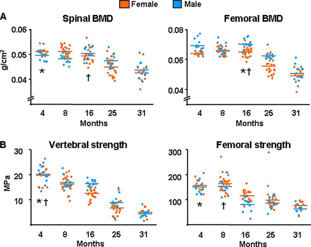
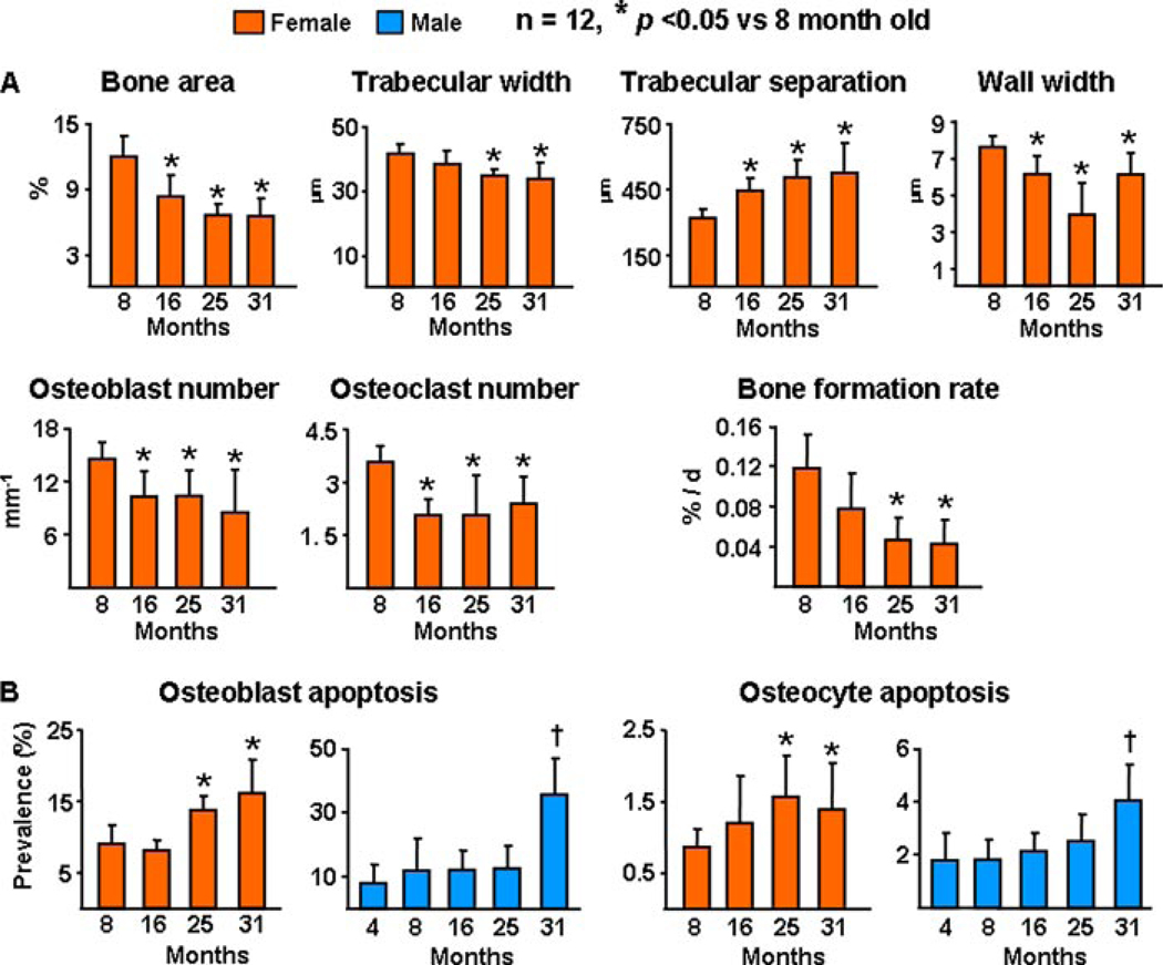
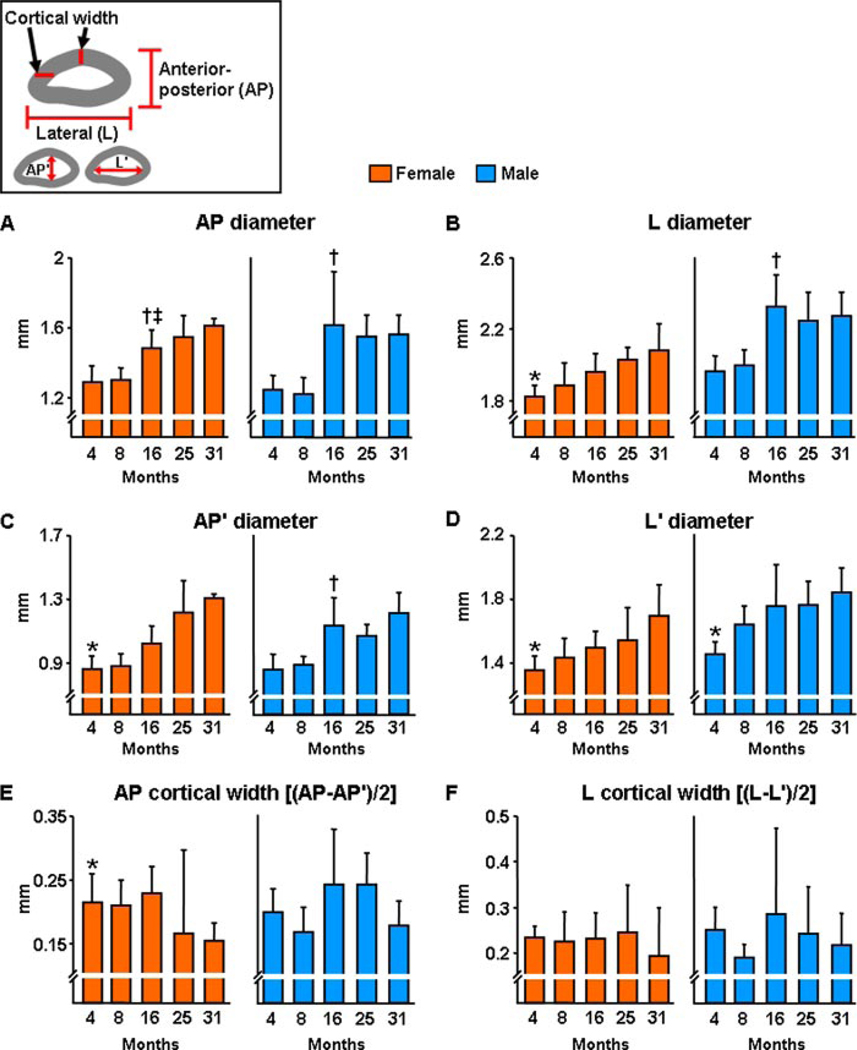
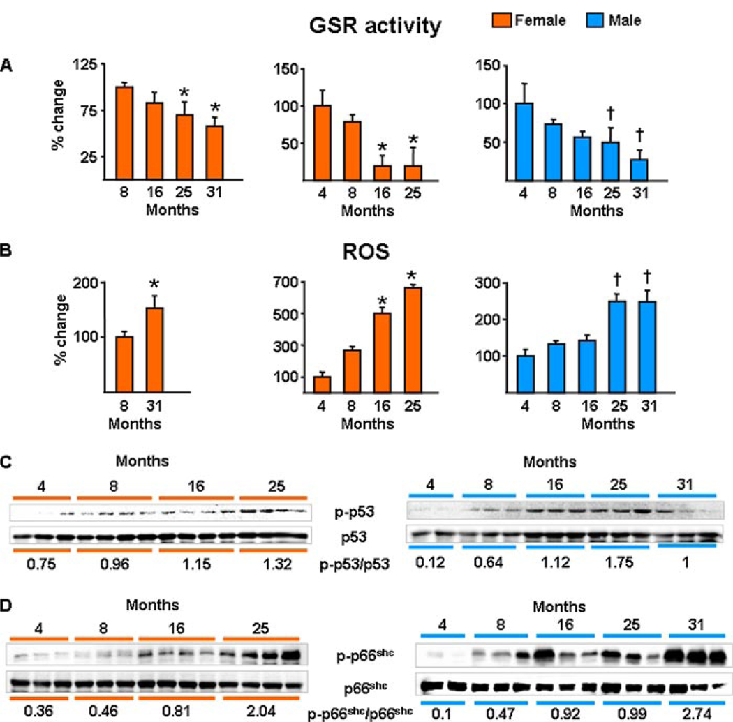
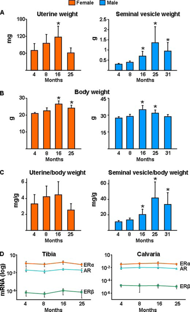
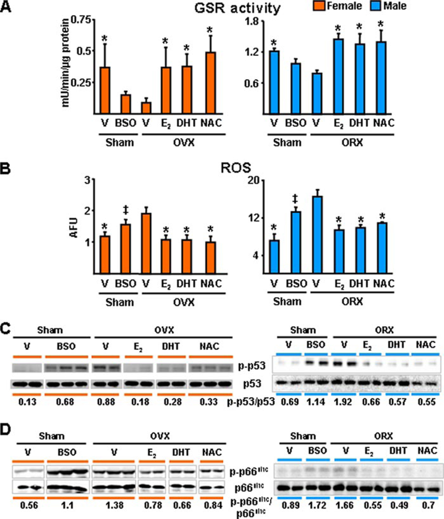
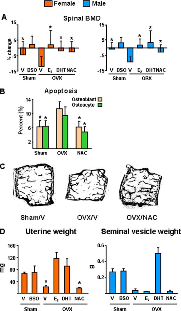
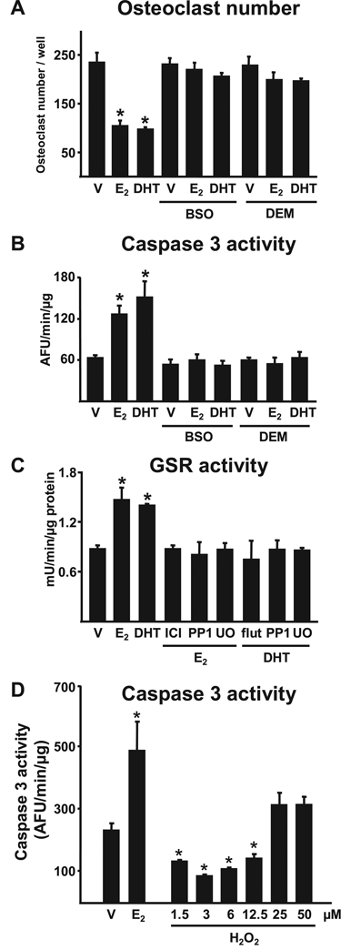
Both aging and loss of sex steroids have adverse effects on skeletal homeostasis, but whether and how they may influence each others negative impact on bone remains unknown. We report herein that both female and male C57BL/6 mice progressively lost strength (as determined by load-to-failure measurements) and bone mineral density in the spine and femur between the ages of 4 and 31 months. These changes were temporally associated with decreased rate of remodeling as evidenced by decreased osteoblast and osteoclast numbers and decreased bone formation rate; as well as increased osteoblast and osteocyte apoptosis, increased reactive oxygen species levels, and decreased glutathione reductase activity and a corresponding increase in the phosphorylation of p53 and p66(shc), two key components of a signaling cascade that are activated by reactive oxygen species and influences apoptosis and lifespan. Exactly the same changes in oxidative stress were acutely reproduced by gonadectomy in 5-month-old females or males and reversed by estrogens or androgens in vivo as well as in vitro. We conclude that the oxidative stress that underlies physiologic organismal aging in mice may be a pivotal pathogenetic mechanism of the age-related bone loss and strength. Loss of estrogens or androgens accelerates the effects of aging on bone by decreasing defense against oxidative stress.


Figures

FIGURE 1
FIGURE 1. BMD and strength decrease with age in sex steroid sufficient female or male C57BL/6 mice
A, BMD at the spine and femur was assessed by dual energy x-ray absorptiometry in two experiments with female and one experiment with male mice. The n was 12 animals per age group in each experiment with females, and 10 – 12 in the experiment with males. In the two experiments with females the age-dependent changes were statistically indistinguishable, hence the data were combined. B, load-to-failure, a measure of strength, was determined by compression testing of the 6th lumbar vertebra (L6) and by 3-point bending of the left femur. Colored horizontal lines in A and B indicate the mean values for each sex. * and † indicate the age after which a time-dependent decline began in females and males, respectively.
FIGURE 2
FIGURE 2. Bone remodeling and bone formation rate decrease with age in sex steroid sufficient female or male C57BL/6 mice, whereas osteoblast and osteocyte apoptosis increases
A, static and dynamic histomorphometric analysis of longitudinal undecalcified sections of L1–L4 vertebrae. Osteoblasts were enumerated on sections from the same specimens stained with toluidine blue and bone formation rate was determined from tetracycline-labeled surfaces. B, osteoblast and osteocyte apoptosis were determined by in situ end-labeling. Bars indicate mean ± S.D.; * or † indicate p < 0.05 versus 8-month-old animals in females and 4-month-old in males. In A, only mice from the first experiment with females were analyzed.
FIGURE 3
FIGURE 3. The external and internal diameters of the femoral diaphysis increase with age in both female and male C57BL/6 mice
The geometry of the left femurs from the two experiments with females and the single experiment with males was determined after performance of the 3-point bending test at the diaphyseal breakpoint by measuring the diameters depicted in the inset. The anterior-posterior external diameter (AP) and lateral exterior diameter (L) were measured using a digital caliper; and the corresponding internal diameters, (AP′) and (L′), were measured with a hand-held microscope, as described under “Experimental Procedures.” † indicates significant difference only between 6 and 18 months by ANOVA. ‡ indicates a progressive increase after 16 months of age. * indicates progressive increase after 4 months of age without significant departure from linearity for the entire data set.
FIGURE 4
FIGURE 4. Oxidative stress increases with age in the bone of female or male C57BL/6 mice
GSR activity (A) and ROS levels (B) were determined in bone marrow aspirates. The results from the two experiments with females and the single experiment with males are shown separately (n = 4 animals per group per experiment). Bars indicate mean ± S.D.; * or † indicate p < 0.05 versus 8-month-old animals in females and 4-month-old in males. C, the levels of phosphorylated p53; and D, p66shc were determined by Western blot analyses in vertebral lysates; each lane represents one animal. The mean ratio of phosphorylated to total protein is depicted numerically at the bottom of the corresponding blots.
FIGURE 5
FIGURE 5. Uterine or seminal vesicle weight or the estrogen and androgen receptor mRNA levels do not change with age in C57BL/6 mice
A, wet uterine or seminal vesicle weight; total body weight (B) and uterine or seminal vesicle (C) corrected for body weight of mice from the second experiment with females and the single experiment with males are shown. Bars represent mean ± S.D.; and n = 10–12 animals per group. * indicates p < 0.05 versus 4-month-old animals. D, mRNA levels of ERα, ERβ, or AR were determined by quantitative PCR in calvaria and tibia obtained from the second experiment with females (n = 5–9), as described under “Experimental Procedures.”
FIGURE 6
FIGURE 6. The antioxidant NAC, as well as estrogens or androgens, prevent gonadectomy-induced increase in oxidative stress in females and males
A–D, 5-month-old mice were sham-operated, OVX, or ORX. Sham-operated animals were administered vehicle (V) or BSO twice a day. One day after surgery, OVX and ORX animals were injected daily with E2 (30 ng/g) or NAC (100 mg/kg) or were implanted with 60-day slow-release pellets containing DHT (10 mg). Animals were sacrificed 6 weeks later. In A and B, n = 4 animals per group. The results depicted for males in D were reproduced in a second blot, in which lysates from two more animals were assayed. The mean ratio of phosphorylated to total protein is depicted numerically in the bottom of the corresponding blots; and, in the case of the male data, represents the results from all four animals. * indicates p < 0.05 versus vehicle-treated OVX or ORX animals; and ‡ indicates p < 0.05 versus sham operated animals treated with vehicle.
FIGURE 7
FIGURE 7. The antioxidant NAC, as well as estrogens or androgens, prevent gonadectomy-induced bone loss and osteoblast and osteocyte apoptosis in females and males
A, spinal BMD was determined by dual energy x-ray absorptiometry 1 to 3 days before and 6 weeks after surgery in the mice of the experiments shown in Fig. 4. The mean ± S.D. of the percent change from the pre-surgery measurement is shown (n = 10–12 per group). B, osteoblast and osteocyte apoptosis were determined in longitudinal undecalcified sections of L1–L4 by in situ end labeling, n = 7–13 per group. * indicates p < 0.05 versus vehicle-treated OVX or ORX animals; and ‡ indicates p < 0.05 versus sham operated animals treated with vehicle. C, representative photomicrographs of lumbar vertebrae from the female mice of the experiment. Note that the loss of central cancellous bone in the OVX/vehicle (OVX/V), as compared with the Sham/vehicle control (Sham/V), has been prevented in the OVX animals that received NAC (OVX/NAC). Unstained and viewed at ×25 with no coverslip. D, wet uterine or seminal vesicle weight of female and male mice. Bars represent mean ± S.D.; and n = 10 –12 animals per group. * indicates p < 0.05 versus vehicle treated sham operated animals.
FIGURE 8
FIGURE 8. Estrogens or androgens regulate osteoclastogenesis and the survival of osteoclasts via antioxidant actions
A and B, bone marrow-derived osteoclasts were treated for 1 h with BSO (10−6 m) or DEM (10−4 m), followed by E2 or DHT (10−8 m) for 24 h. Osteoclasts were enumerated after staining for TRAPase and apoptosis was quantified by determining caspase 3 activity. C, GSR activity in osteoclasts treated with ICI 182,780 (10−7 m), flutamide (10−7 m), PP1 (10−6 m), or U0126 (10−6 m) for 1 h followed by the indicated steroids for 24 h. D, apoptosis was quantified by determining caspase 3 activity in bone marrow-derived osteoclasts treated for 24 h with 10−8 m E2 or the indicated doses of H2O2. Bars indicate mean ± S.D. of triplicate determinations; * p < 0.05 versus vehicle.
FIGURE 9
FIGURE 9. Estrogens or androgens regulate the survival of osteoblasts via antioxidant actions
A, apoptosis was quantified by determining caspase 3 activity in OB-6 cells treated for 1 h with BSO or DEM followed by the steroids for 1 and 6 h with the pro-apoptotic agent etoposide (5 × 10⁻⁵ m), TNFα (10−9 m), or H2O2 (5 × 10⁻⁵ m). Bars indicate mean ± S.D. of triplicate determinations; * indicates p < 0.05 versus vehicle. B, C2C12 cells transfected with a vector control or wild-type p66shc plasmid, along with green fluorescent protein, were treated with or without H2O2 (5 × 10⁻⁵ m). The number of apoptotic cells was determined by examining the nuclear morphology of fluorescent cells 6 h later. Bars indicate mean ± S.D. of triplicate determinations; * p < 0.05 versus vector control untreated. C, OB-6 cells were treated for 1 h with the MEK inhibitor PD98059 (5 × 10⁻⁵ m), then the indicated steroids (10−8 m) were added, and 1 h later the cultures were exposed for 15 min to H2O2 (5 × 10⁻⁵ m). Phosphorylated p66shc was determined by Western blot analyses in cell lysates.
FIGURE 10
FIGURE 10. ROS-activated signals affecting the genesis and lifespan of osteoblasts and osteoclasts and the counter-regulatory actions of sex steroids
Effects of ROS, exemplified here by H2O2, on the genesis and survival of both osteoblasts and osteoclasts are depicted in black, and as shown require the same signaling cascades and factors, i.e. ERKs, NF-κB, and osteoclastogenic cytokines like Rankl, Tnf, and interleukin (IL) 6, used by estrogens to regulate the birth and death of osteoblasts and osteoclasts, albeit in the exactly opposite manner (13, 14). Like most other cell types, bone cells attempt to counteract the adverse effects of ROS by several defense mechanisms (depicted in blue). Such mechanisms include the up-regulation of ROS scavenging enzymes (superoxide dismutases and catalases) as well as DNA-damage repair genes by Forkhead transcription factors (FoxO; see accompanying article, Almeida et al. (82)). Additionally, enzymes like glutathione peroxidase use glutathione to reduce ROS to alcohols. Glutathione reductase is a key partner in this cycle because it converts the disulfide (GSSG) back into glutathione (GSH). The pro-apoptotic effects of H2O2 on osteoblasts (and probably their mesenchymal stem cell progenitors) are associated with phosphorylation of p53 and p66shc. ROS decreases osteoblastogenesis by at least two mechanisms: 1) antagonism of Wnt signaling by diversion of β-catenin from Tcf- to FoxO-mediated transcription (see accompanying article, Almeida et al. (82)); and 2) direct and sustained activation of ERKs and NF-κB (78). ROS inhibit osteoclast apoptosis and stimulate osteoclastogenesis by increasing RANKL production in cells of the stromal/osteoblastic lineage (79) as well as an ERK/NF-κB/Tnf/interleukin 6-mediated mechanism(39).E2 or DHT antagonizes the effects of ROS via several mechanisms(depicted in red): (a) up-regulation of GSR(and thioredoxin reductase activity (39, 80); (b) attenuation of p66shc phosphorylation via a Src- and ERK-dependent pathway; and (c) down-regulation of the production of osteoclastogenic cytokines like TNF and interleukin 6 via attenuation of NF-κB. In addition, E2 or DHT antagonizes the effects of ROS by attenuating osteoblast apoptosis and stimulating osteoclast apoptosis via a transient and sustained ERK activation, respectively; and by inhibiting osteoclastogenesis, through a sustained ERK activation (81). For the interplay between ROS and estrogens on osteoblastogenesis, see “Discussion.”

Similar articles

Cited by

References

    1. Looker AC, Wahner HW, Dunn WL, Calvo MS, Harris TB, Heyse SP, Johnston CC, Jr, Lindsay R. Osteoporosis Int. 1998;8:468–489. - PubMed
    1. Hui SL, Slemenda CW, Johnston CC., Jr J. Clin. Investig. 1988;81:1804–1809. - PMC - PubMed
    1. Kleerekoper M, Villanueva AR, Stanciu J, Rao DS, Parfitt AM. Calcif. Tissue Int. 1985;37:594–597. - PubMed
    1. Burr DB, Turner CH, Naick P, Forwood MR, Ambrosius W, Hasan MS, Pidaparti R. J. Biomech. 1998;31:337–345. - PubMed
    1. Heaney RP. Bone. 2003;33:457–465. - PubMed