. 2007 Aug 1;120(Pt 15):2532-43.
doi: 10.1242/jcs.009878. Epub 2007 Jul 10.

GNL3L inhibits activity of estrogen-related receptor gamma by competing for coactivator binding

Affiliations

GNL3L inhibits activity of estrogen-related receptor gamma by competing for coactivator binding

Hiroaki Yasumoto et al. J Cell Sci. .

Abstract

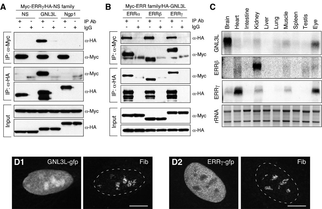
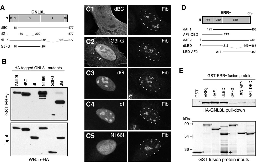
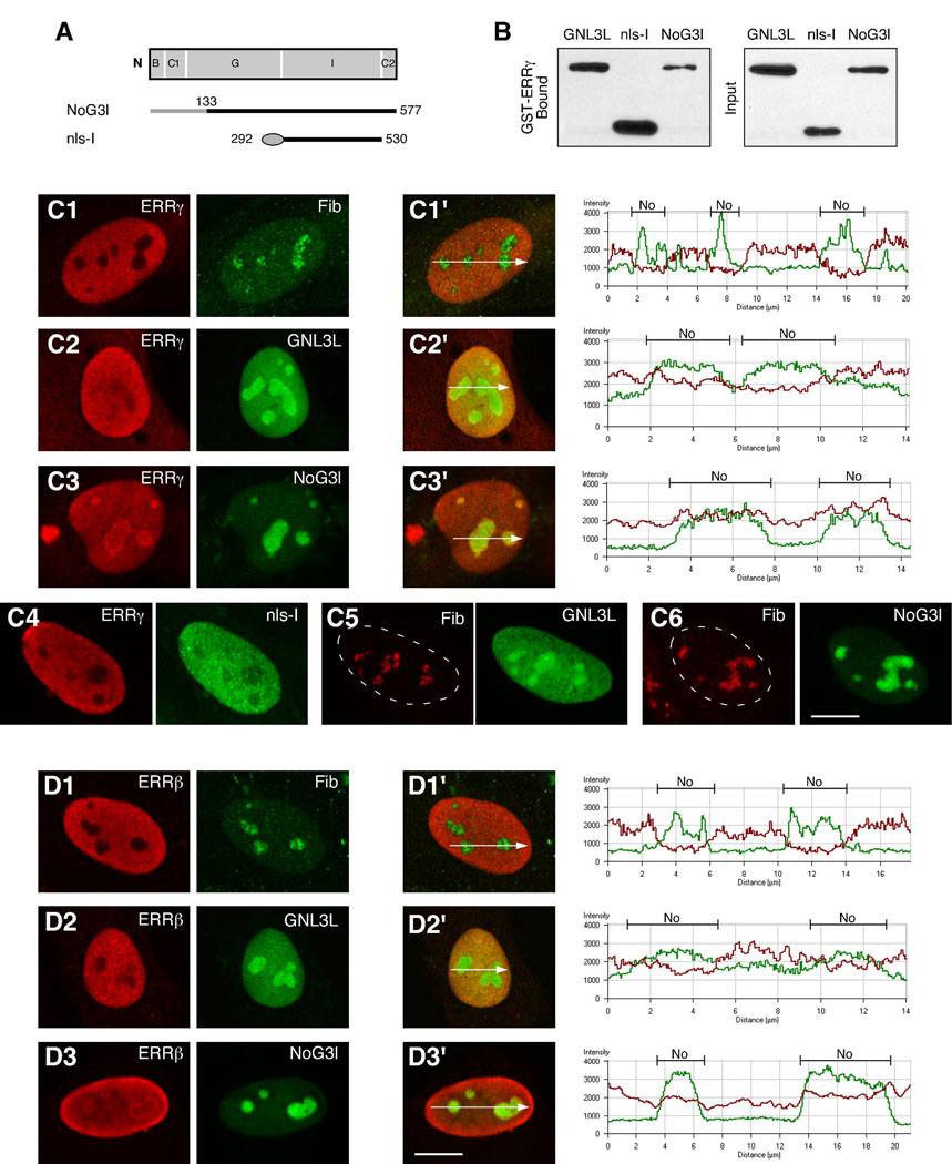
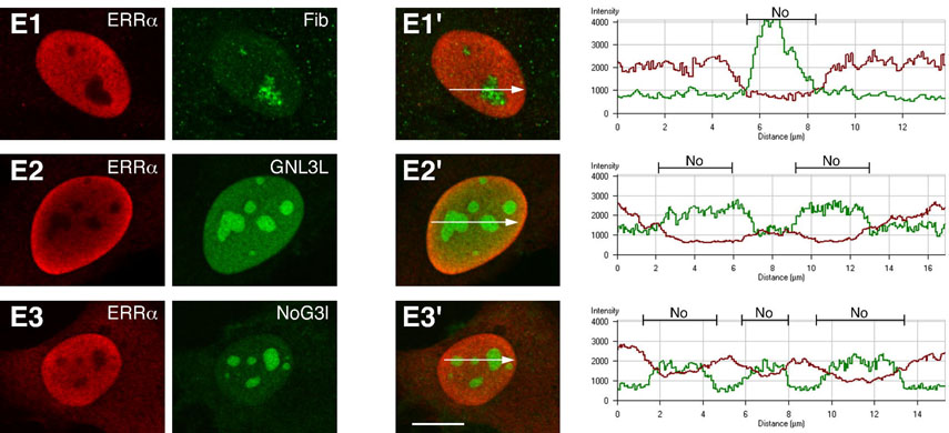
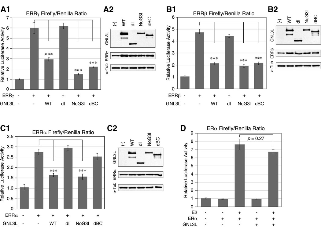
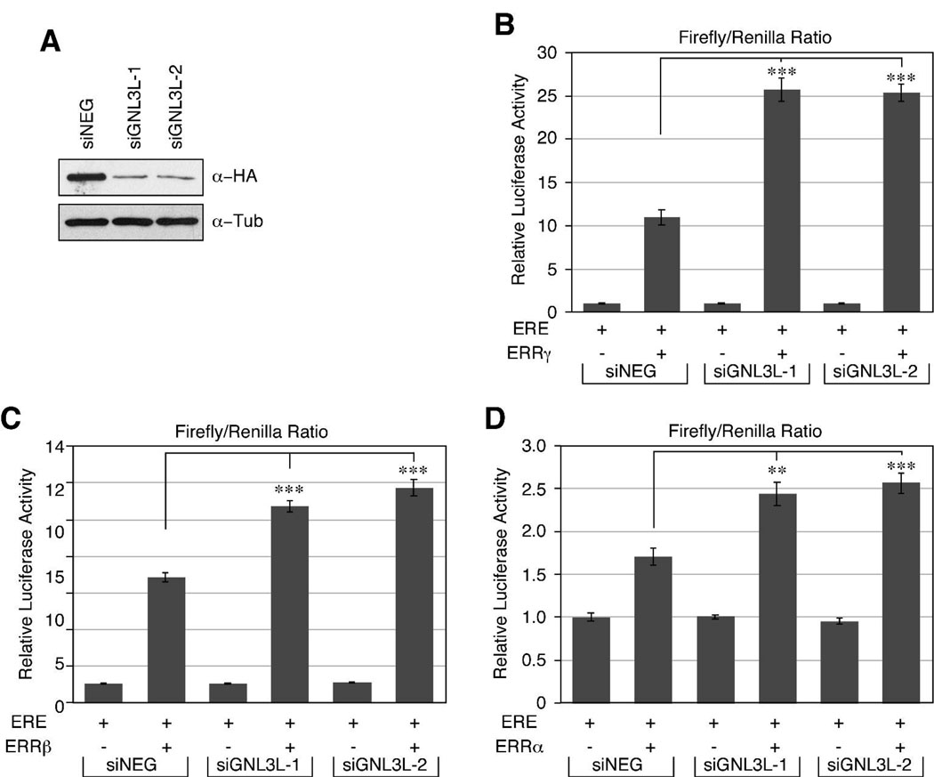
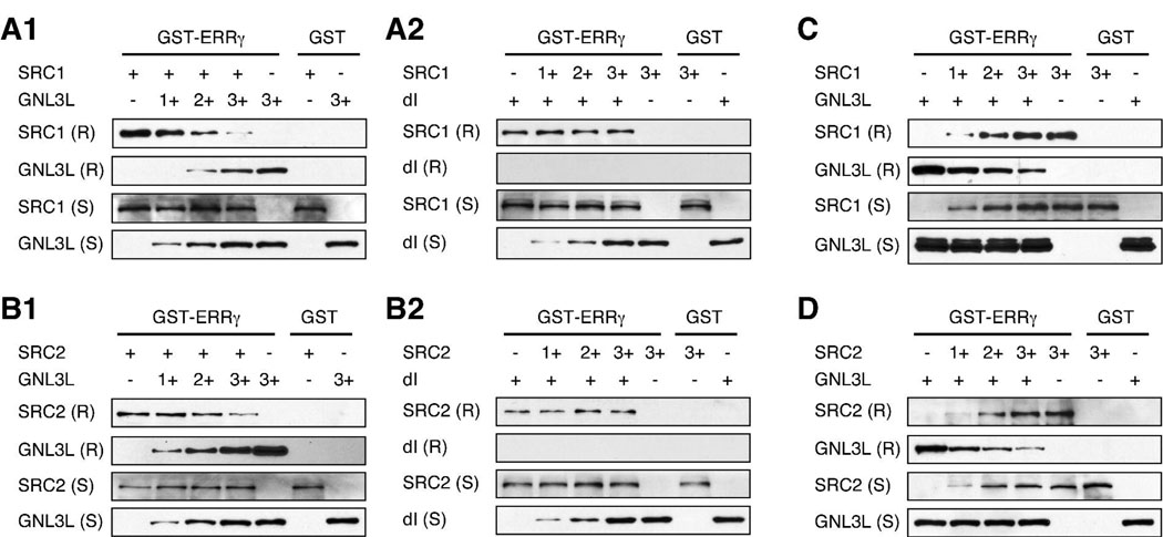
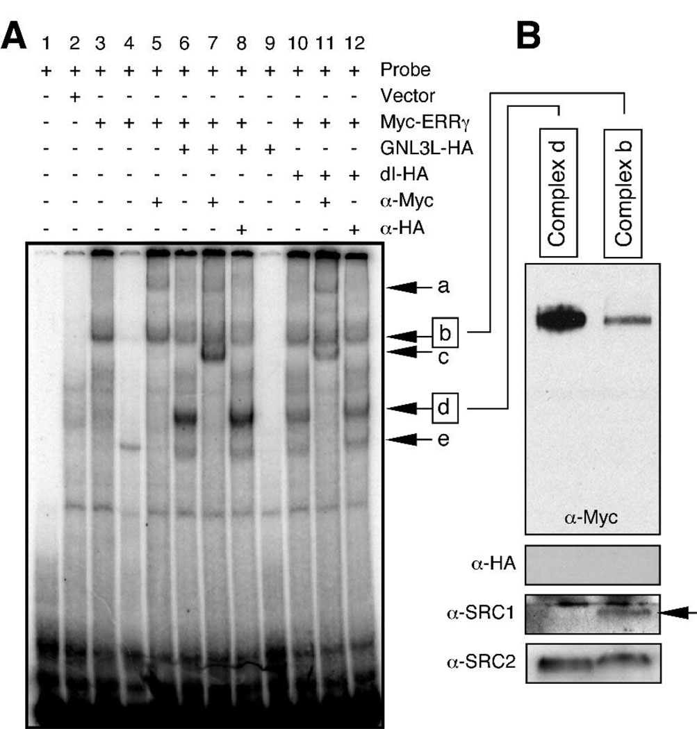
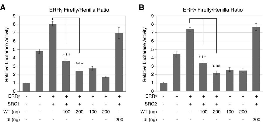
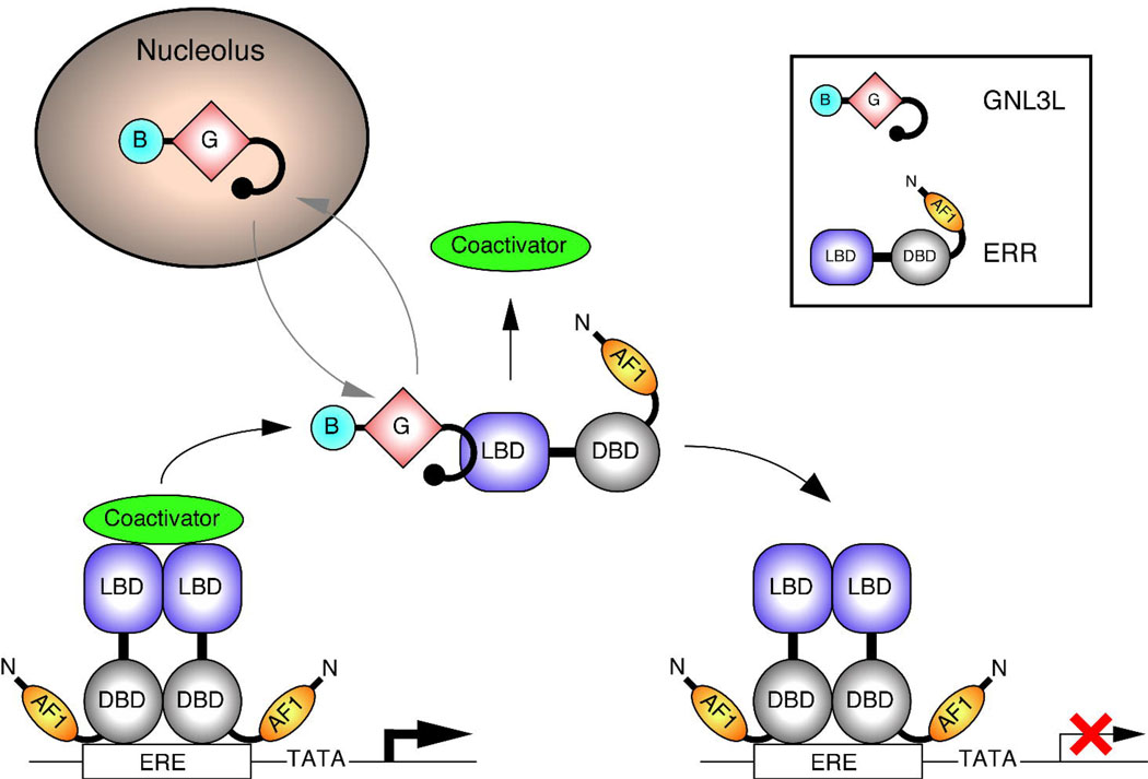
Guanine nucleotide binding protein-like 3 (GNL3L) is the closest homologue of a stem cell-enriched factor nucleostemin in vertebrates. They share the same yeast orthologue, Grn1p, but only GNL3L can rescue the growth-deficient phenotype in Grn1-null yeasts. To determine the unique function of GNL3L, we identified estrogen-related receptor gamma (ERRgamma) as a GNL3L-specific binding protein. GNL3L and ERRgamma are coexpressed in the eye, kidney and muscle, and co-reside in the nucleoplasm. The interaction between GNL3L and ERRgamma requires the intermediate domain of GNL3L and the AF2-domain of ERRgamma. Gain-of- and loss-of-function experiments show that GNL3L can inhibit the transcriptional activities of ERR genes in a cell-based reporter system, which does not require the nucleolar localization of GNL3L. We further demonstrate that GNL3L is able to reduce the steroid receptor coactivator (SRC) binding and the SRC-mediated transcriptional coactivation of ERRgamma. This work reveals a novel mechanism that negatively regulates the transcriptional function of ERRgamma by GNL3L through coactivator competition.


Figures

Figure 1
Figure 1. GNL3L interacts with estrogen receptor-related proteins (ERR)-α, β, and γ
Protein interactions between ERRγ and nucleostemin family genes (A) and between GNL3L and ERR family genes (B) were examined by in vivo coimmunoprecipitation assays. HEK293 cells were cotransfected with: (A) Myc-tagged ERRγ and HA-tagged nucleostemin, GNL3L, or Ngp1 expression plasmids, or (B) HA-tagged GNL3L and Myc-tagged ERRα, β, or γ expression plasmids. Lysates were immunoprecipitated (IP) with anti-Myc antibody (rows 1 and 2, α-Myc) or anti-HA antibody (rows 3 and 4, α-HA). The copurified proteins (rows 1 and 3) and the immunoprecipitates (rows 2 and 4) were immunodetected with the antibodies indicated on the right. Our results show that ERRγ interacts with only GNL3L, but not with nucleostemin or Ngp1, and that GNL3L binds all ERR family proteins. (C) Tissue distributions of GNL3L, ERRβ, and ERRγ in adult mice are shown by multi-tissue northern blots. The GNL3L message is expressed primarily in the neural tissues, including the brain and eye, and at a lower level in the kidney and muscle. The expression levels of GNL3L and ERRγ match in the kidney, muscle, and eye, but differ in the brain and heart. (D) In U2OS cells, the intensity of the green fluorescent protein (GFP)-tagged GNL3L (GNL3L-gfp) is higher in the nucleolus than in the nucleoplasm. GFP-tagged ERRγ (ERRγ-gfp) is localized exclusively in the nucleoplasm. The nucleolar regions are labeled by anti-fibrillarin (Fib) immunofluorescence in the right panels. Dashed lines demarcate the nucleocytoplasmic boundaries. Bars: 10 um.
Figure 2
Figure 2. Binding between GNL3L and ERRγ requires the intermediate (I)-domain of GNL3L and the AF2-domain of ERRγ
(A) Truncated mutants of GNL3L were used to determine its interacting domain with ERRγ. B, basic; C1 and C2, coiled-coil domain-1 and 2; G, GTP-binding domain; I, intermediate domain. Numbers indicate amino acid positions. (B) GST-ERRγ fusion proteins fail to bind mutants that lack the I-domain (dI and G3l-G), but can retain the dBC and the non-GTP-binding mutants, N166I and dG. (C) The subcellular distributions of HA-tagged dBC, G3l-G, dG, dI, and N166I mutants were shown by confocal analyses double-labeled with anti-HA (left panels) and anti-fibrillarin (Fib, right panels) antibodies. Bar: 10um. (D) Truncated mutants of ERRγ were used to determine its interacting domain with GNL3L. AF1 and AF2, activation function 1 and 2; DBD, DNA-binding domain; LBD, ligand-binding domain. (E) Affinity binding assays show that GST fusion proteins of the wild-type ERRγ, the dAF1 mutant, and the dLBD mutant can bind GNL3L, but GST fusion proteins of the dAF2, LBD-AF2, and AF1-DBD mutants cannot (top panel). The amounts of GST fusion proteins used in each reaction, marked by asterisks, are shown in the bottom panel by Commassie blue staining. Some degradation occurs at the fusion site of the GST-dLBD protein (arrow).
Figure 3
Figure 3. Overexpression of GNL3L brings ERRβ and ERRγ into the nucleolus
(A) To generate a nucleolar form of GNL3L (NoG3l), we replaced the N-terminal nucleolus-targeting domain of GNL3L with the corresponding region of nucleostemin (grey bar), which has a stronger nucleolus-targeting activity than GNL3L but lacks the ability to bind ERRγ. To create a nucleoplasmic form of GNL3L (nls-I), we fused the I-domain of GNL3L with an SV40 nuclear localization signal (oval). (B) Affinity binding assays show that both nls-I and NoG3l maintain the ability to bind ERRγ. To measure the effect of GNL3L overexpression on the distribution of ERRγ, U2OS cells were transfected with Myc-tagged ERRγ alone (C1), Myc-tagged ERRγ and HA-tagged wild-type or mutant GNL3L (C2–4), or HA-tagged GNL3L constructs alone (C5–6). Double-transfected cells were labeled with anti-Myc (red) and anti-HA (green) immunofluorescence, and visualized by confocal analyses. Single-transfected cells were immunostained with anti-fibrillarin antibody (Fib) and anti-Myc or anti-HA antibody. The ERRγ (red) fluorescence intensities are measured quantitatively along the lines indicated by arrows, shown in the right panels of (C1’–C3’), and the nucleolar regions (No) are indicated by the increase of green fluorescence. Compared to cells transfected with only ERRγ, the fluorescence intensity of ERRγ in the nucleolus is increased in cells cotransfected with NoG3l or the wild-type GNL3L. In contrast, the nls-I mutant does not change the distribution of ERRγ (C4). Neither does ERRγ alter the distribution of GNL3L (C5) or NoG3l (C6). The same analyses were performed using ERRβ (D1–D3) and ERRα (E1–E3). Our results showed that when coexpressed with wild-type GNL3L (D2) or NoG3l (D3), the ERRβ signals begin to accumulate in the nucleolus. GNL3L overexpression has little or no effect on the distribution of ERRα (E2–E3). Bars: 10um.
Figure 4
Figure 4. Overexpression of GNL3L inhibits the transcriptional activities of ERR proteins independent of nucleolar distribution
(A1) Estrogen response element (ERE)-specific transcriptional activities were measured in CV-1 cells by the ratio between the ERE-driven Firefly luciferase activity and the Renilla-null luciferase activity. ERRγ elicits a six-fold increase in the ERE-specific transcriptional activity. Coexpression of wild-type GNL3L (WT) leads to a 50% reduction in the ERRγ-mediated transcriptional activity. This decrease is reversed by a deletion of the ERRγ-binding I-domain of GNL3L (dI). Coexpression of either the nucleolar form (NoG3l) or the nucleoplasmic form (dBC) of GNL3L suppresses the ERRγ transcriptional activity more than or to the same extent as the wild-type GNL3L protein. Using the same approach, we show that this inhibitory activity of GNL3L can also work on ERRβ (B1) and ERRα (C1) with the exception that the dBC mutant has little effect on the ERRα-mediated transactivation. Error bars represent stand error of mean (s.e.m.). ***, P value < 0.0001. (A2, B2, C2) The expression levels of wild-type and mutant GNL3L proteins and ERR proteins in the experimental samples are compared side-by-side by anti-HA and anti-Myc western blots, respectively. Anti-α-tubulin western blots (α-Tub) are used as loading controls. (D) GNL3L fails to suppress the estradiol (E2)-induced transcriptional activity of ERα on the ERE-driven promoter in the same cell-based reporter system.
Figure 5
Figure 5. The endogenous GNL3L suppresses the transcriptional activities of ERR family genes
(A) To confirm the GNL3L-mediated negative regulation of ERR activities from a loss-of-function angle, a short interference RNA (siRNA) approach was used to knock down the endogenous expression of GNL3L. Compared to the control knockdown sample (siNEG), the protein knockdown efficiencies of GNL3L-specific siRNA duplexes, siGNL3L-1 and siGNL3L-2, in HEK293 cells stably expressing HA-tagged GNL3L are estimated to be 83% and 84%, respectively. (B) Consistent with our overexpression data, the transcriptional activity of ERRγ is increased 2.5 times by the siGNL3L-1 and siGNL3L-2 treatments as compared to the siNEG-treated sample. (C, D) GNL3L knockdown has the same effect on the ERRβ and ERRα-mediated transactivation, although their increases are less dramatic than the increase in the ERRγ-mediated transactivation. Error bars represent standard error of mean (s.e.m.). **, P value < 0.001; ***, P value < 0.0001.
Figure 6
Figure 6. GNL3L competes with SRC1 and SRC2 for ERRγ binding
Agarose-bound GST fusion proteins of ERRγ (1ug) were used to pull down whole cell lysates containing a fixed amount of SRC1 (A) or SRC2 (B), mixed with increasing amounts of the wild-type GNL3L (A1, B1) or the dI mutant lacking the ERRγ-interacting domain (A2, B2). Whole cell proteins in each sample were adjusted to the same amount. In the agarose-retained portions (R), the interaction between GNL3L and ERRγ can reduce the amount of SRC1 and SRC2 bound by ERRγ in a dose-dependent manner, but the dI mutant fails to do so. Conversely, when GST-ERRγ fusion proteins were used to pull down the same amount of GNL3L in the presence of increasing amounts of SRC1 (C) or SRC2 (D), SRC1 and SRC2 were able to reduce the amount of GNL3L bound by ERRγ in a dose-dependent way as well. Proteins in the agarose-bound fraction and in the supernatant are indicated by (R) and (S), respectively.
Figure 7
Figure 7. Coexpression of GNL3L increases the electrophoretic mobility of the DNA-bound ERRγ protein complex and reduces its binding with SRC1 and SRC2
(A) The GNL3L effect on the DNA binding of ERRγ was examined by electrophoretic mobility shift assays (EMSA) using ERE-containing probes and whole cell lysates expressing the indicated recombinant proteins. Compared to the probe alone (lane 1) and the vector-transfected control sample (lane 2), the ERRγ-specific DNA-protein complex can be identified in lane 3 (arrow b), competed by excess non-labeled probes (lane 4), and supershifted by anti-Myc antibody (lane 5, arrow a). Coexpression of GNL3L produces fast-moving complexes (lane 6, arrows d and e), which can be supershifted by anti-Myc antibody (lane 7, arrow c) but not by anti-HA antibody (lane 8). GNL3L itself cannot bind the ERE probe (lane 9). The intensity of the fast-moving complex d is reduced by a deletion of the ERRγ-binding I-domain of GNL3L (lanes 10–12). (B) The fast-moving complex d and the slow-moving complex b were retrieved from the EMSA gel, fractionated in SDS-denaturing PAGE, and analyzed for their ERRγ (α-Myc), GNL3L (α-HA), SRC1, and SRC2 protein components by western blottings. Our results indicate that the increase in the electrophoretic mobility of the ERRγ-DNA complex by GNL3L coexpression can be explained by a loss of SRC1 binding (arrow) and diminished SRC2 binding, rather than by protein cleavage of ERRγ.
Figure 8
Figure 8. GNL3L suppresses SRC-mediated transcriptional coactivation of ERRγ
(A) Using the same cell-based reporter system as described in Fig. 4, we show that the ERE-specific transcriptional activity in cells coexpressing ERRγ and SRC1 (8.0±0.3) is 1.7 times higher than that of the ERRγ-expressing sample (4.8±0.3). When coexpressed with the wild-type GNL3L (WT), this ERRγ and SRC1-mediated ERE-specific transcriptional activity is reduced by 55 and 70 percent compared to the sample expressing both ERRγ and SRC1 in a dose-dependent manner. This inhibitory effect of GNL3L on the SRC1-mediated coactivation of ERRγ requires the I-domain of GNL3L, as a deletion of this domain (dI) fails to suppress the transcriptional activity of ERRγ and SRC1 (P value = 0.17). (B) Using the same approach, we show that GNL3L can also suppress the coactivator function of SRC2 on the ERRγ-dependent transcriptional activity in a dose-dependent (54% reduction for 100ng of GNL3L and 71% reduction for 200ng of GNL3L) and I-domain-dependent (P value = 0.58) manner. Error bars represent stand error of mean (s.e.m.). ***, P value < 0.0001.
Figure 9
Figure 9. GNL3L inhibits the transcriptional activities of ERR family genes by coactivator competition
Our data reveal a novel mechanism that regulates the activity of ERR family genes by a nucleolar GTP-binding protein GNL3L. GNL3L decreases the transcriptional activity of ERR proteins. This event takes place in the nucleoplasm and does not require the nucleolar localization of GNL3L. The interaction between GNL3L and ERRγ displaces coactivators such as SRC1 and SRC2 from the ERRγ complex. The SRC-depleted ERRγ protein binds DNA without GNL3L, resulting in transcriptional inhibition. In this model, the nucleolar accumulation of GNL3L does not appear to affect its ability to suppress the transcriptional function of ERR proteins (grey arrows). Abbreviations for protein domains of GNL3L and ERR are explained in Fig. 2A and 2D.

Similar articles

Cited by

References

    1. Ariazi EA, Jordan VC. Estrogen-related receptors as emerging targets in cancer and metabolic disorders. Curr Top Med Chem. 2006;6:203–215. - PubMed
    1. Baddoo M, Hill K, Wilkinson R, Gaupp D, Hughes C, Kopen GC, Phinney DG. Characterization of mesenchymal stem cells isolated from murine bone marrow by negative selection. J Cell Biochem. 2003;89:1235–1249. - PubMed
    1. Beekman C, Nichane M, De Clercq S, Maetens M, Floss T, Wurst W, Bellefroid E, Marine JC. Evolutionarily Conserved Role of Nucleostemin: Controlling Proliferation of Stem/Progenitor Cells during Early Vertebrate Development. Mol Cell Biol. 2006;26:9291–9301. - PMC - PubMed
    1. Bonnelye E, Vanacker JM, Spruyt N, Alric S, Fournier B, Desbiens X, Laudet V. Expression of the estrogen-related receptor 1 (ERR-1) orphan receptor during mouse development. Mech Dev. 1997;65:71–85. - PubMed
    1. Bookout AL, Jeong Y, Downes M, Yu RT, Evans RM, Mangelsdorf DJ. Anatomical profiling of nuclear receptor expression reveals a hierarchical transcriptional network. Cell. 2006;126:789–799. - PMC - PubMed

Publication types

MeSH terms