. 2007 Jul 17;104(29):12099-104.
doi: 10.1073/pnas.0705268104. Epub 2007 Jul 10.

IL-6-dependent and -independent pathways in the development of interleukin 17-producing T helper cells

Affiliations

IL-6-dependent and -independent pathways in the development of interleukin 17-producing T helper cells

Akihiro Kimura et al. Proc Natl Acad Sci U S A. .

Abstract

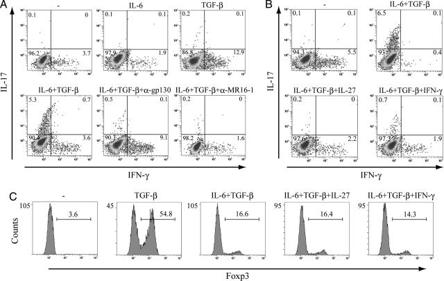
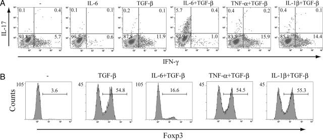
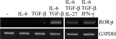
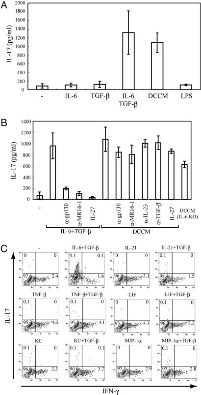
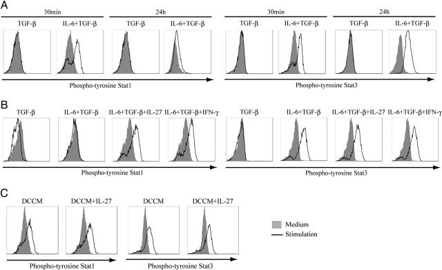
CD4(+) T cells producing IL-17 [T helper (Th)17], as distinct from Th1 or Th2 cells, have recently been shown to be associated with autoimmunity, but it is not entirely clear how Th17 cells are generated from naïve T cells. We demonstrate here that IL-6, but not TNF-alpha or IL-1beta, can, in combination with TGF-beta, induce Th17 cell generation from naïve T cells and inhibit TGF-beta-induced Foxp3 expression. Moreover, conditioned medium from lipopolysaccharide-stimulated bone marrow-derived dendritic cells (DCCM) can induce IL-17 production in naïve T cells. Interestingly, IL-17 was produced by DCCM even with the addition of anti-gp130 antibody or DCCM from IL-6 KO mice. The combination of IL-6 and TGF-beta could maintain activation of signal transducer and activator of transcription (Stat)3, but not of Stat1. IL-27 or IFN-gamma suppressed the induction of Th17 cells by TGF-beta plus IL-6 and maintained Stat1 activation under these conditions. In contrast, both Stat1 and Stat3 remained to be activated in naïve T cells cultured with DCCM. These findings represent a different basis for Th17 differentiation from naïve T cells.


Conflict of interest statement

The authors declare no conflict of interest.

Figures

Fig. 1.
Fig. 1.
The combination of IL-6 and TGF-β induces the development of Th17 cells. Isolated naïve T cells were cultured with anti-CD3/CD28 beads and the indicated cytokines for 3 days. (A and B) Naïve T cells were stimulated with the indicated cytokines in the presence or absence of anti-mouse gp130 antibody or anti-mIL-6R monoclonal antibody MR16-1. After stimulation, cells were restimulated with PMA and ionomycin for 5 h and with GolgiStop (last 2 h) and were then subjected to intracellular cytokine staining. Dot plots show intracellular staining for IFN-γ and IL-17. (C) Foxp3 expression was determined by staining with anti-mouse Foxp3 antibody. These results are representative of three independent experiments.
Fig. 2.
Fig. 2.
TNF-α or IL-1β in combination with TGF-β has no effect on the differentiation of Th17 cells. (A) MACS-sorted naïve T cells were cultured with anti-CD3/CD28 beads. Cells were stimulated with IL-6 or TGF-β alone and with IL-6, TNF-α, or IL-1β combined with TGF-β. After 3 days, cells were restimulated with PMA and ionomycin before intracellular cytokine staining for IFN-γ and IL-17 and analysis by flow cytometry. Dot plots show intracellular staining for IFN-γ and IL-17. (B) Isolated naïve T cells were activated with anti-CD3/CD28 beads and cultured with TGF-β alone and with IL-6, TNF-α, or IL-1β combined with TGF-β. Foxp3 expression was determined by staining with anti-mouse Foxp3 antibody. These results are representative of three independent experiments.
Fig. 3.
Fig. 3.
RORγt mRNA expression by IL-6 and TGF-β with or without IL-27 or IFN-γ. Isolated naïve T cells were stimulated with anti-CD3/CD28 beads and the indicated cytokines, followed by total RNA and cDNA prepared as described in Materials and Methods. RORγt induction was examined by using RT-PCR.
Fig. 4.
Fig. 4.
IL-17 is produced by DCCM without IL-6-mediated activity in naïve T cells. (A) Purified naïve T cells were cultured with anti-CD3/CD28 beads and the indicated cytokines, LPS, or CM from DCs stimulated with LPS. (B) Naïve T cells were stimulated with TGF-β plus IL-6, DCCM, or DCCM from IL-6 KO mice in the presence or absence of IL-27 or neutralizing antibodies for IL-6. Supernatants were collected 2 days after stimulation, and IL-17 production was measured by means of ELISA. Data show means ± SE of three independent experiments. (C) MACS-sorted naïve T cells were activated with anti-CD3/CD28 beads. Cells were cultured with the indicated stimulators. After 3 days, cells were restimulated with PMA and ionomycin before intracellular cytokine staining for IFN-γ and IL-17 and analysis by flow cytometry. Dot plots show intracellular staining for IFN-γ and IL-17.
Fig. 5.
Fig. 5.
Activation of Stat1 and Stat3 by stimulation to produce IL-17 in naïve T cells. MACS-sorted naïve T cells were cultured with anti-CD3/CD28 beads. (A) Naïve T cells were stimulated with TGF-β alone or TGF-β plus IL-6 for 30 min or 24 h. (B) Naïve T cells were stimulated with TGF-β alone or TGF-β plus IL-6 in the presence or absence of IL-27 or IFN-γ for 24 h. (C) Naïve T cells were cultured with DCCM with or without IL-27. Cells were fixed and permeabilized in 90% methanol, followed by staining with Alexa Fluor 488-conjugated phospho-Stat1 and phycoerythrin-conjugated phospho-Stat3. Intracellular phospho-Stat1 and -Stat3 were measured by flow cytometry. These results are representative of three independent experiments.

Similar articles

Cited by

References

    1. Fontenot JD, Rasmussen JP, Williams LM, Dooley JL, Farr AG, Rudensky AY. Immunity. 2005;22:329–341. - PubMed
    1. Szabo SJ, Kim ST, Costa GL, Zhang X, Fathman CG, Glimcher LH. Cell. 2000;100:655–669. - PubMed
    1. Zheng W, Flavell RA. Cell. 1997;89:587–596. - PubMed
    1. Hsieh CS, Macatonia SE, Tripp CS, Wolf SF, O'Garra A, Murphy KM. Science. 1993;260:547–549. - PubMed
    1. Scharton TM, Scott P. J Exp Med. 1993;178:567–577. - PMC - PubMed

Publication types

MeSH terms