Review
. 2007 Oct;28(4):161-78.
doi: 10.1016/j.yfrne.2007.05.003. Epub 2007 Jun 8.

Topography in the preoptic region: differential regulation of appetitive and consummatory male sexual behaviors

Affiliations
Review

Topography in the preoptic region: differential regulation of appetitive and consummatory male sexual behaviors

Jacques Balthazart et al. Front Neuroendocrinol. 2007 Oct.

Abstract

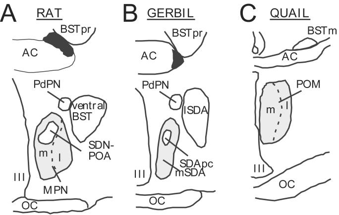
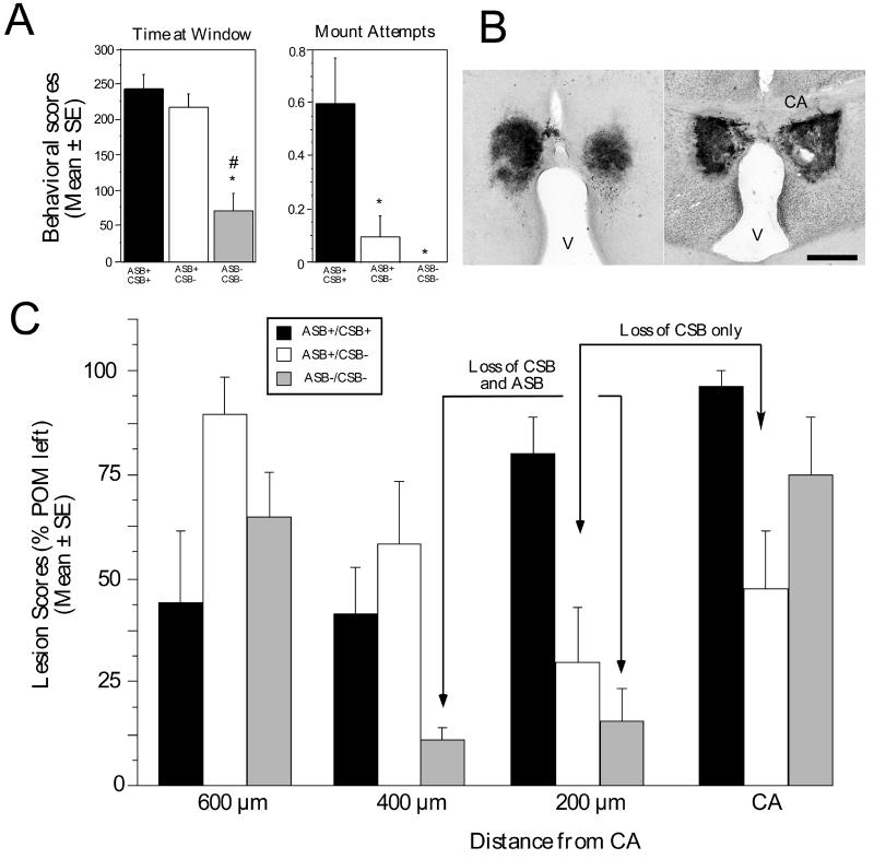
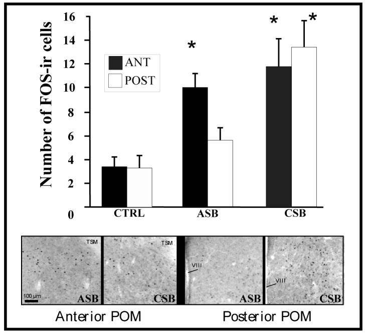
Several studies have suggested dissociations between neural circuits underlying the expression of appetitive (e.g., courtship behavior) and consummatory components (i.e., copulatory behavior) of vertebrate male sexual behavior. The medial preoptic area (mPOA) clearly controls the expression of male copulation but, according to a number of experiments, is not necessarily implicated in the expression of appetitive sexual behavior. In rats for example, lesions to the mPOA eliminate male-typical copulatory behavior but have more subtle or no obvious effects on measures of sexual motivation. Rats with such lesions still pursue and attempt to mount females. They also acquire and perform learned instrumental responses to gain access to females. However, recent lesions studies and measures of the expression of the immediate early gene c-fos demonstrate that, in quail, sub-regions of the mPOA, in particular of its sexually dimorphic component the medial preoptic nucleus, can be specifically linked with either the expression of appetitive or consummatory sexual behavior. In particular more rostral regions can be linked to appetitive components while more caudal regions are involved in consummatory behavior. This functional sub-region variation is associated with neurochemical and hodological specializations (i.e., differences in chemical phenotype of the cells or in their connectivity), especially those related to the actions of androgens in relation to the activation of male sexual behavior, that are also present in rodents and other species. It could thus reflect general principles about POA organization and function in the vertebrate brain.


Figures

Figure 1
Figure 1
Schematic dimorphic nuclei identified in the rat, gerbil and Japanese quail brain. The homologies between rat and gerbil are presented according to the recent work of Finn and Yahr [69] and panels A and B are redrawn from a figure published in that study. The quail medial preoptic nucleus (POM) appears to be homologous to the MPN of rats based on arguments that are presented in the text. Abbreviations: AC: anterior commissure; BST: bed nucleus of the stria terminalis; BSTm: bed nucleus of the stria terminalis, medial part; BSTpr: bed nucleus of the stria terminalis principal nucleus; l: lateral; lSDA: lateral part of the sexually dimorphic area; m: medial; MPN: medial preoptic nucleus (in rats); mSDA: ; OC: optic chiasma; PdPN: posterodorsal preoptic nucleus; POM: medial preoptic nucleus (in quail); SDApc: sexually dimorphic area, pars compacta; SDN-POA: sexually dimorphic nucleus of the preoptic area; III: third ventricle.
Figure 2
Figure 2
In Japanese quail, the medial preoptic nucleus (POM) is sexually dimorphic and its boundaries are specifically outlined by a dense cluster of aromatase-immunoreactive (ARO-ir) cells. The volume of POM and the number of ARO-ir cells in this nucleus represent a signature of testosterone action in the brain. A. Schematic presentation of the anatomical localization of the POM, which extends through most of the rostro-caudal extent of the preoptic area. At a level just caudal to the anterior commissure (CA), the POM merges with the medial part of the bed nucleus of the stria terminalis (BSTm). The distribution of ARO-ir cells in this brain area (illustrated in gray tone) overlaps exactly with the boundaries of POM and BSTm. Sections are arranged in a rostral to caudal order from top to bottom. B. Photomicrograph illustrating a section through the preoptic area at the level of the anterior commissure that was stained by immunocytochemistry for aromatase. The two groups of ARO-ir cells outlining the POM and BSTm are clearly visible. C. Photomicrographs of the left POM stained by immunocytochemistry for aromatase respectively in a sexually mature gonadally intact male (Intact), in a castrated male (CX) and in a castrated male treated with testosterone (CX+T). D. Quantification of the ARO-ir cells in the POM. In the entire POM, the number of ARO-ir cells is decreased by castration and restored by a treatment with exogenous testosterone (left graph; redrawn from [75]). Separate counts reveal that effects of castration and testosterone are more prominent in the lateral than in the medial part of the nucleus (right graph; redrawn from [12]). Note that data in the two parts of this panel come from different studies in which quantification was performed by different methods in sample fields of different sizes. Absolute numbers of ARO-ir cells thus cannot be directly compared and attention should be paid only to relative changes induced by castration and testosterone treatment. Magnification bar in B-C= 500 μm. Abbreviations: AM: nucleus anterior medialis hypothalami; BSTl: bed nucleus of the stria terminalis, lateral part; BSTm: bed nucleus of the stria terminalis, medial part; CA: commissura anterior; E: entopallium; FPL: lateral forebrain bundle; GLv: nucleus geniculatus lateralis, pars ventralis; HA: hyperpallium apicale; Hp: hippocampus; LA: nucleus lateralis anterior thalami; N: nidopallium; nCPa; POM: nucleus preopticus medialis; SL: nucleus septalis lateralis; SM: nucleus sepatalis medialis; TSM: tractus septopallio-mesencephalicus; V: third ventricle; VLT: nucleus ventrolateralis thalami.
Figure 3
Figure 3
Stereotaxic implants of testosterone in the POM activate male copulatory behavior in male Japanese quail. A. Schematic drawings illustrating the position of implants that did or did not activate male-typical sexual behavior. Implants restore copulatory behavior in castrated male quail only if located within the limits of the POM but not in adjacent structures of the medial preoptic area. The panel shows two coronal sections located at the level of the anterior commissure (CA: bottom) and 180 μm more rostrally (top). Filled circles represent implants that restored at least mount attempts in castrates, open circles represent implants that were behaviorally ineffective. Implants that are only 100-200 μm lateral to POM are ineffective. AC: nucleus accumbens (now renamed nucleus striae terminalis lateralis; see [113]); CA: commissura anterior; FPL: fasciculus prosencephali lateralis; POM: nucleus preopticus medialis; SL: nucleus septalis lateralis; SM: nucleus septalis medialis. Redrawn from [22]. B. Testosterone implants in the POM also induce the expression of aromatase that can be detected by the quantification of the numbers of ARO-ir cells on the implantation side. The intensity of sexual behavior activated by the implants (frequency of mount attempts during the last two behavioral tests before sacrifice) correlates best with the induction of aromatase expression (numbers of ARO-ir cells) in sections located just rostrally (100-200 μm) to the anterior commissure (CA). The figure represents the correlations (Pearson's product-moment coefficient) between individual measures of sexual behavior and numbers of ARO-ir cells in the left or right POM (counted in one 20 μm-thick section every 100 μm). The testosterone implant was always positioned on the right side. Coefficients that are significantly different from zero are indicated by an asterisk (Redrawn from [23]). C-D. Photomicrographs illustrating the POM as observed in sections stained by immunocytochemistry for aromatase in two castrated quail that received a stereotaxic implant filled with cholesterol (C) or with testosterone (D) in the dorso-lateral part of the nucleus on the right side of the brain. The induction of aromatase by testosterone as evidenced by an increase in the number of ARO-ir cells in clearly visible in panel D. The tip of the implants is clearly visible and labeled by an asterisk. The dotted line indicates the limits of the POM. Magnification bar= 500 μm (modified from [23]).
Figure 4
Figure 4
Aromatase-immunoreactive cells located in the medial preoptic nucleus (POM) send dense projection the premotor mesencephalic periacqueductal gray (PAG), this projection is denser in males than in females and this sex difference is more prominent in the caudal than in the rostral part of the nucleus. Retrogradely labeled cells are also more numerous in the lateral than in the medial part of the nucleus A. Schematic representation of the POM-PAG projection and of the experimental design used to study this connection. A retrograde tracer (fluorescent latex beads) was injected in PAG, retrogradely transported to POM neurons that project to PAG and then identified in sections through the POM that were also stained by immunocytochemistry for aromatase. B. Representative photomicrographs through the lateral (left) or medial (right) POM illustrating the presence of retrogradely transported latex beads (red) that had been injected in PAG in aromatase-immunoreactive cells (green). In the medial POM a large fraction of ARO-ir cells project to the PAG, this proportion is smaller in the lateral POM. Arrows point to ARO-ir cells containing red latex bed that project to the PAG, arrowheads indicate ARO-ir cells that do not project to PAG C. Number (mean±SEM) of retrogradely fluorescent (RLF) aromataseimmunoreactive (ARO-ir) A cells counted in 4 different locations in the POM (medial and lateral field at a rostral and caudal level) in male (black bars) and female (hatched bars) quail illustrating the effects of the position within POM (medial vs. lateral and rostral vs. caudal) on the number of ARO-RLF cells and the presence of larger numbers in males as compared to females. Post hoc MANOVA confirmed that the sex difference is significant only in the lateral caudal POM (*=p<0.05) but statistical tendencies (p<0.10) are also observed in the medial POM at both rostrocaudal levels. Redrawn from [48].
Figure 5
Figure 5
Electrolytic lesions affecting the POM just rostral to the anterior commissure inhibit male consummatory sexual behavior (CSB), more rostral lesions also inhibit appetitive sexual behavior (ASB). A. Behavioral scores (time at window during the learned social proximity test [ASB] or mount attempt frequencies [CSB]) computed for three subgroups of birds bearing a POM lesion defined by the presence of appetitive sexual behavior and a low level of consummatory sexual behavior (ASB+/CSB+), or by the complete absence of consummatory behavior associated or not with an inhibition of appetitive sexual behavior (ASB/CSB− and ASB+/CSB− respectively). *=p < 0.05 by comparison with ASB+/CSB+ subgroup ; #=p < 0.05 by comparison with ASB+/CSB− subgroup). B. Photomicrographs of sections stained by immunocytochemistry for aromatase illustrating a bilateral electrolytic lesion in the rostral part of the POM (left) and in the caudal part of the nucleus at the level of the anterior commissure (CA). Magnification bar= 500 μm, V: third ventricle. C. Average lesions scores observed in the POM at four rostro-caudal levels (identified by reference to the commissural anterior, CA) in the three subgroups of birds defined by their behavior as described in panel A. Lesions were scored on a 5 point scale with 0= no POM left (complete lesion) and 4= 100 % of POM left (no lesion at this level). A nearly complete loss of CSB is observed in birds bearing large lesions at the CA level or 200 μm more rostrally. The most important deficits in ASB are observed in birds bearing large lesions 200 or 400 μm rostral to CA. Redrawn from data in [29].
Figure 6
Figure 6
Performance of consummatory sexual behavior (CSB) induces FOS expression throughout the medial preoptic nucleus (POM) while performance of appetitive sexual behavior (ASB; production rhythmic cloacal sphincter movements in response to the view of a female) induces FOS in the anterior POM only. The bar graph represents the number of FOSimmunoreactive cells counted in sections through the anterior (ANT) or posterior (POST) POM in birds that had expressed either ASB or CSB 90 min before sacrifice or had been handled as a control (CTRL) manipulation. *= significantly different from the control situation. The lower part of the panel shows photomicrographs through representative sections in the ASB and CSB groups. Redrawn from data in [137].

Similar articles

Cited by

References

    1. Absil P, Riters LV, Balthazart J. Preoptic aromatase cells project to the mesencephalic central gray in the male Japanese quail (Coturnix japonica) Horm. Behav. 2001 - PubMed
    1. Absil P, Braquenier JB, Balthazart J, Ball GF. Effects of lesions of nucleus taeniae on appetitive and consummatory aspects of male sexual behavior in Japanese quail. Brain Behav. Evol. 2002;60:13–35. - PubMed
    1. Adkins EK. Hormonal basis of sexual differentiation in the Japanese quail. J. Comp. Physiol. Psychol. 1975;89:61–71. - PubMed
    1. Adkins EK, Boop JJ, Koutnik DL, Morris JB, Pniewski EE. Further evidence that androgen aromatization is essential for the activation of copulation in male quail. Physiol. Behav. 1980;24:441–446. - PubMed
    1. Adkins-Regan E. Hormones and animal social behavior. Princeton University Press; Princeton, NJ: 2005. p. 411.

Publication types