. 2007:3:125.
doi: 10.1038/msb4100165. Epub 2007 Jul 10.

Nano-enabled synthetic biology

Affiliations

Nano-enabled synthetic biology

Mitchel J Doktycz et al. Mol Syst Biol. 2007.

Abstract

Biological systems display a functional diversity, density and efficiency that make them a paradigm for synthetic systems. In natural systems, the cell is the elemental unit and efforts to emulate cells, their components, and organization have relied primarily on the use of bioorganic materials. Impressive advances have been made towards assembling simple genetic systems within cellular scale containers. These biological system assembly efforts are particularly instructive, as we gain command over the directed synthesis and assembly of synthetic nanoscale structures. Advances in nanoscale fabrication, assembly, and characterization are providing the tools and materials for characterizing and emulating the smallest scale features of biology. Further, they are revealing unique physical properties that emerge at the nanoscale. Realizing these properties in useful ways will require attention to the assembly of these nanoscale components. Attention to systems biology principles can lead to the practical development of nanoscale technologies with possible realization of synthetic systems with cell-like complexity. In turn, useful tools for interpreting biological complexity and for interfacing to biological processes will result.


Figures

Figure 1
Figure 1
Overview of a genetic-based synthetic cell. The membrane of synthetic cells can be created from a variety of materials, including natural membrane components, synthetic polymers, and micro- or nano-fabricated materials. Alternatively, a water-in-oil emulsion is used to define the ‘cell'. Typical cell volumes are on the order of picoliters to nanoliters and contain genetic material and appropriate transcription and translation machinery. The incorporation of pore structures into the membrane facilitates transfer of appropriate reagents.
Figure 2
Figure 2
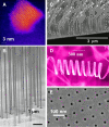
Collage of synthetic nanoscale materials. (A) 0-d nanoscale material shown in a Z-STEM image of 655 QdotsTM (CdSe/CdS core/shell, Quantum Dot Corporation; image courtesy of Professor S Rosenthal, Vanderbilt University (adapted with permission from McBride et al, 2006)). (BD) 1-d nanoscale materials: (B) silicon nanowires grown on a silicon wafer (image courtesy of Professsor P Yang, University of California, Berkeley (adapted with permission from Goldberger et al, 2006)); (C) carbon nanofibers with orientation controlled by an electrical field that was switched during the growth process from 90 to 45° with respect to the plane of the substrate (Merkulov et al, 2002c); (D) ZnO nanospring resulting from a polar surface-induced growth phenomena (Image courtesy Professor ZL Wang, Georgia Tech (adapted with permission from Kong and Wang, 2003)). (E) A 50-nm thick silicon nitride membrane (2-d material) with a regular array of 25-nm diameter holes (image courtesy of Dr HD Tong, Nanosens, and Dr HV Jansen, University of Twente (adapted with permission from Tong et al, 2004)).
Figure 3
Figure 3
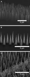
Micrographs of carbon nanofibers. (A) Vertically aligned carbon nanofibers can be prepared from a continuous catalyst stripe yielding an array of CNFs that are randomly arranged. (B) Individual CNFs can be precisely positioned using electron beam lithography to define catalyst sites for growth of individual nanofibers (Melechko et al, 2005). (C) These patterning techniques can be combined on a single substrate to yield both ordered and disorder arrays of CNFs.
Figure 4
Figure 4
Fabrication of a cell mimic array. The device is created using (A) contact photolithography and ICP-RIE etching of silicon to define a fluidic channel approximately 10 μm in depth. (B, C) Metal lift-off of Ni catalyst is followed by PECVD growth of CNF forests that rise slightly above the channel ceiling. (D) Structures are sealed with a PDMS lid, following selective ink-jet introduction of reagents (15 pl) into mimic cells, as diagrammed in the upper right and seen in the fluorescent micrograph at bottom left. The image at bottom right shows the structure after sealing and wetting of the device. The materials are contained, but fluid diffuses between the chambers (Fletcher et al, 2004).

References

    1. Aharoni A, Amitai G, Bernath K, Magdassi S, Tawfik DS (2005a) High-throughput screening of enzyme libraries: Thiolactonases evolved by fluorescence-activated sorting of single cells in emulsion compartments. Chem Biol 12: 1281–1289 - PubMed
    1. Aharoni A, Griffiths AD, Tawfik DS (2005b) High-throughput screens and selections of enzyme-encoding genes. Curr Opin Chem Biol 9: 210–216 - PubMed
    1. Ajayan PM, Ebbesen TW (1997) Nanometre-size tubes of carbon. Rep Prog Phys 60: 1025–1062
    1. Anderson PW (1972) More is different—broken symmetry and nature of hierarchical structure of science. Science 177: 393–396 - PubMed
    1. Andrianantoandro E, Basu S, Karig DK, Weiss R (2006) Synthetic biology: new engineering rules for an emerging discipline. Mol Syst Biol 2: 2006.002816738572 - PMC - PubMed

Publication types