

Comparison between ultrasound and noncontrast helical computed tomography for identification of acute ureterolithiasis in a teaching hospital setting
- PMID: 17625708
- PMCID: PMC11014692
- DOI: 10.1590/s1516-31802007000200007
Comparison between ultrasound and noncontrast helical computed tomography for identification of acute ureterolithiasis in a teaching hospital setting
Abstract
Context and objective: Recent studies have shown noncontrast computed tomography (NCT) to be more effective than ultrasound (US) for imaging acute ureterolithiasis. However, to our knowledge, there are few studies directly comparing these techniques in an emergency teaching hospital setting. The objectives of this study were to compare the diagnostic accuracy of US and NCT performed by senior radiology residents for diagnosing acute ureterolithiasis; and to assess interobserver agreement on tomography interpretations by residents and experienced abdominal radiologists.
Design and setting: Prospective study of 52 consecutive patients, who underwent both US and NCT within an interval of eight hours, at Hospital São Paulo.
Methods: US scans were performed by senior residents and read by experienced radiologists. NCT scan images were read by senior residents, and subsequently by three abdominal radiologists. The interobserver variability was assessed using the kappa statistic.
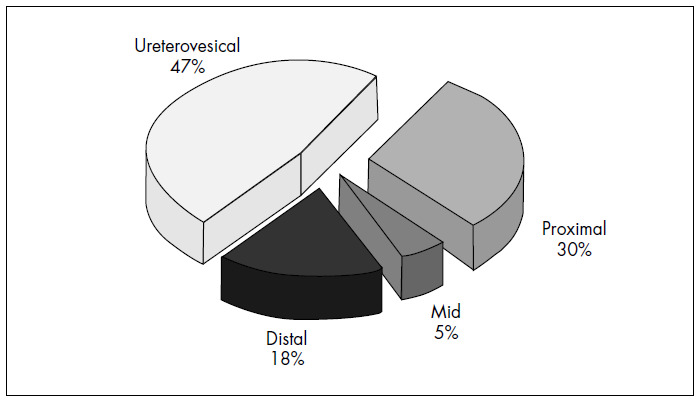
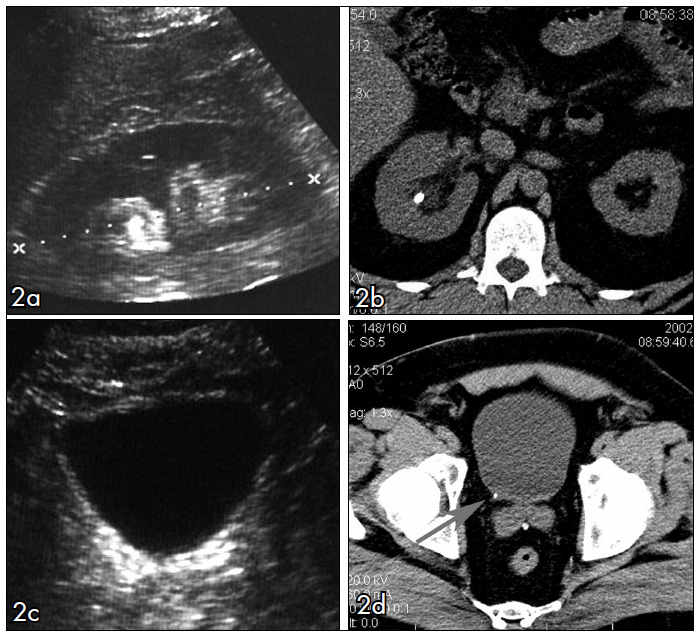
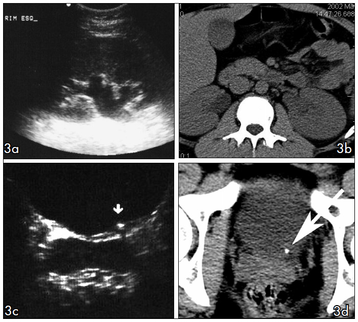
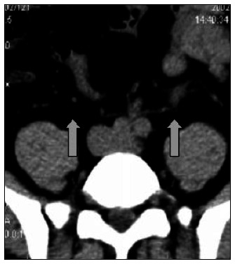
Results: Ureteral calculi were found in 40 out of 52 patients (77%). US presented sensitivity of 22% and specificity of 100%. When collecting system dilatation was associated, US demonstrated 73% sensitivity, 82% specificity. The interobserver agreement in NCT analysis was very high with regard to identification of calculi, collecting system dilatation and stranding of perinephric fat.
Conclusions: US has limited value for identifying ureteral calculi in comparison with NCT, even when collecting system dilatation is present. Residents and abdominal radiologists demonstrated excellent agreement rates for ureteral calculi, identification of collecting system dilatation and stranding of perinephric fat on NCT.
CONTEXTO E OBJETIVO:: Estudos atuais demonstram que a tomografia computadorizada helicoidal sem contraste (TC) apresenta maior acurácia do que a ultra-sonografia (US) no diagnóstico da ureterolitíase aguda, porém, poucos estudos a esse respeito foram realizados em atendimento radiológico de urgência de hospital universitário. Nossos objetivos foram comparar a sensibilidade diagnóstica da US com a TC realizadas por residentes no diagnóstico de ureterolitíase aguda e comparar a análise da TC interpretada por residentes e radiologistas experientes.
TIPO DE ESTUDO E LOCAL:: Estudo prospectivo de 52 pacientes com cólica renal aguda, que foram submetidos a exame de US seguido de TC em período máximo de oito horas no Hospital São Paulo.
MÉTODOS:: Os exames de US foram realizados por médicos residentes e conferidos pelos preceptores, já os de TC foram analisados por outro residente e posteriormente analisados por três radiologistas independentes.
RESULTADOS:: Nos 52 pacientes analisados foram encontrados 40 cálculos ureterais na TC (77%). A US apresentou uma sensibilidade de 22% e especificidade de 100%, que aumentou para 73% e 82% respectivamente, quando se associou a identificação da dilatação do sistema coletor. A TC analisada pelo residente e pelos radiologistas apresentou uma excelente correlação para identificação do cálculo ureteral, para heterogeneidade da gordura peri-renal e para dilatação do sistema coletor.
CONCLUSÕES:: A US realizada pelos residentes tem menor sensibilidade no diagnóstico da litíase ureteral, quando comparada à TC, mesmo quando associada à presença de dilatação do sistema coletor. Residentes e radiologistas especialistas apresentaram excelente concordância no diagnóstico de litíase ureteral.
Conflict of interest statement
Figures
Similar articles
-
Low-dose and standard-dose unenhanced helical computed tomography for the assessment of acute renal colic: prospective comparative study.Acta Radiol. 2005 Nov;46(7):756-63. doi: 10.1080/02841850500216004. Acta Radiol. 2005. PMID: 16372698
-
Assessment of the clinical utility of the rim and comet-tail signs in differentiating ureteral stones from phleboliths.AJR Am J Roentgenol. 2001 Dec;177(6):1285-91. doi: 10.2214/ajr.177.6.1771285. AJR Am J Roentgenol. 2001. PMID: 11717067
-
Unenhanced helical CT of ureterolithiasis: value of the tissue rim sign.AJR Am J Roentgenol. 1997 Apr;168(4):997-1000. doi: 10.2214/ajr.168.4.9124157. AJR Am J Roentgenol. 1997. PMID: 9124157
-
[Unenhanced spiral CT in urolithiasis: indication, performance and interpretation].Rofo. 2003 Jul;175(7):904-10. doi: 10.1055/s-2003-40426. Rofo. 2003. PMID: 12847644 Review. German.
-
[Diagnosis of ureteral calculi using ultrasonography, intravenous urography and unenhanced helical computed tomography].Med Pregl. 2005 Sep-Oct;58(9-10):503-6. doi: 10.2298/mpns0510503g. Med Pregl. 2005. PMID: 16526256 Review. Serbian.
Cited by
-
Ureterolithiasis and the quest for rational use of diagnostic imaging methods.Radiol Bras. 2018 Nov-Dec;51(6):VII-VIII. doi: 10.1590/0100-3984.2018.51.6e2. Radiol Bras. 2018. PMID: 30559568 Free PMC article. No abstract available.
-
B-mode ultrasound versus color Doppler twinkling artifact in detecting kidney stones.J Endourol. 2013 Feb;27(2):149-53. doi: 10.1089/end.2012.0430. Epub 2013 Jan 30. J Endourol. 2013. PMID: 23067207 Free PMC article. Clinical Trial.
-
Focused ultrasonic propulsion of kidney stones: review and update of preclinical technology.J Endourol. 2013 Oct;27(10):1183-6. doi: 10.1089/end.2013.0315. Epub 2013 Sep 14. J Endourol. 2013. PMID: 23883117 Free PMC article. Review.
-
A systematic review and meta-analysis of clinical signs, symptoms, and imaging findings in patients with suspected renal colic.J Am Coll Emerg Physicians Open. 2022 Dec 1;3(6):e12831. doi: 10.1002/emp2.12831. eCollection 2022 Dec. J Am Coll Emerg Physicians Open. 2022. PMID: 36474707 Free PMC article.
References
-
- Smith RC, Rosenfield AT, Choe KA, et al. Acute flank pain: comparison of non-contrast-enhanced CT and intravenous urography. Radiology. 1995;194(3):789–794. - PubMed
-
- Tamm EP, Silverman PM, Shuman WP. Evaluation of the patient with flank pain and possible ureteral calculus. Radiology. 2003;228(2):319–329. - PubMed
-
- Sourtzis S, Thibeau JF, Damry N, Raslan A, Vandendris M, Bellemans M. Radiologic investigation of renal colic: unenhanced helical CT compared with excretory urography. AJR Am J Roentgenol. 1999;172(6):1491–1494. - PubMed
-
- Goldman SM, Faintuch S, Ajzen SA, et al. Diagnostic value of attenuation measurements of the kidney on unenhanced helical CT of obstructive ureterolithiasis. AJR Am J Roentgenol. 2004;182(5):1251–1254. - PubMed
-
- Lanoue MZ, Mindell HJ. The use of unenhanced helical CT to evaluate suspected renal colic. AJR Am J Roentgenol. 1997;169(6):1579–1584. - PubMed
Publication types
MeSH terms
LinkOut - more resources
Full Text Sources
