. 2007 Sep 14;282(37):26746-26753.
doi: 10.1074/jbc.M705200200. Epub 2007 Jul 10.

Vascular endothelial growth factor (VEGF)-induced up-regulation of CCN1 in osteoblasts mediates proangiogenic activities in endothelial cells and promotes fracture healing

Affiliations

Vascular endothelial growth factor (VEGF)-induced up-regulation of CCN1 in osteoblasts mediates proangiogenic activities in endothelial cells and promotes fracture healing

Athanasios N Athanasopoulos et al. J Biol Chem. .

Abstract

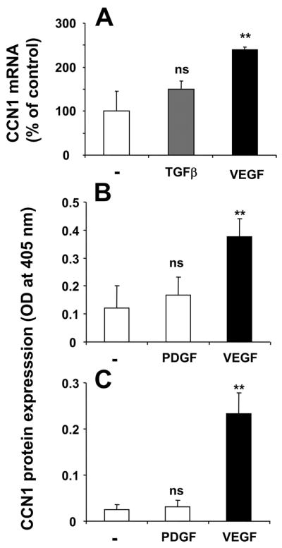
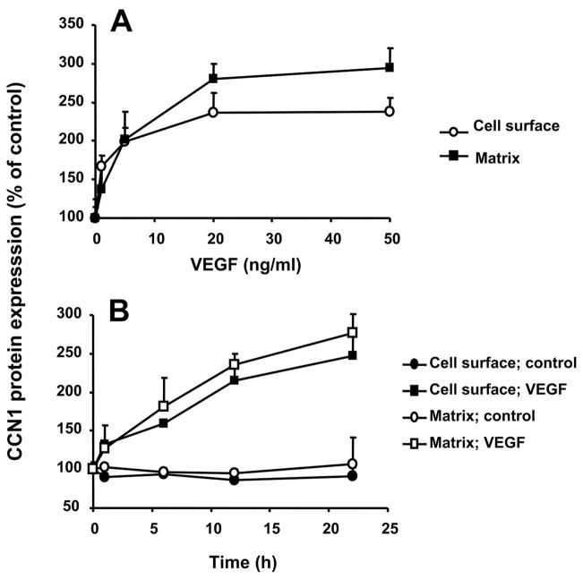
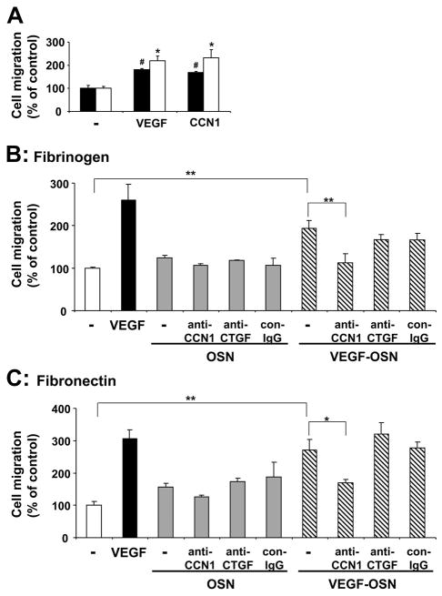
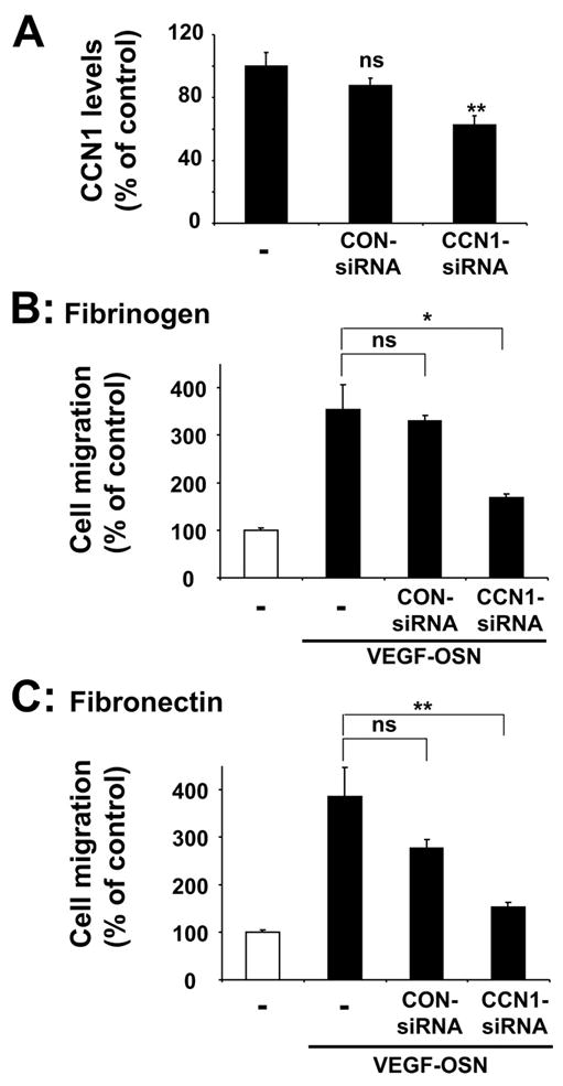
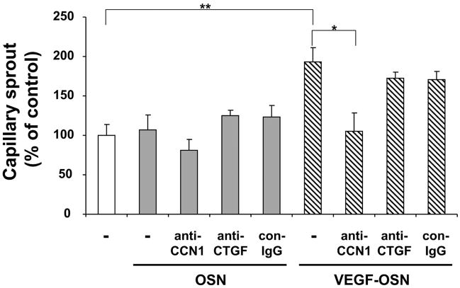
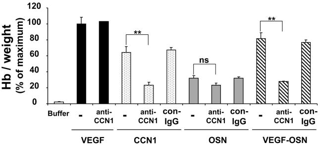
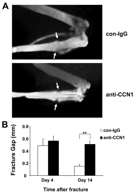
Angiogenesis is indispensable during fracture repair, and vascular endothelial growth factor (VEGF) is critical in this process. CCN1 (CYR61) is an extracellular matrix signaling molecule that has been implicated in neovascularization through its interactions with several endothelial integrin receptors. CCN1 has been shown to be up-regulated during the reparative phase of fracture healing; however, the role of CCN1 therein remains unclear. Here, the regulation of CCN1 expression in osteoblasts and the functional consequences thereof were studied. Stimulation of osteoblasts with VEGF resulted in a dose- and time-dependent up-regulation of CCN1 mRNA and protein. An up-regulation of both cell surface-associated CCN1 as well as extracellular matrix-associated CCN1 in osteoblasts was found. The supernatant of VEGF-prestimulated osteoblasts was chemotactic for endothelial cells, increasing their migration and stimulated capillary-like sprout formation. These effects could be attributed to the presence of CCN1 in the osteoblast supernatant as they were prevented by an antibody against CCN1 or by small interfering RNA-mediated knockdown of osteoblast CCN1. Moreover, the supernatant of VEGF-prestimulated osteoblasts induced angiogenesis in Matrigel plugs in vivo in a CCN1-dependent manner. In addition, blockade of CCN1 prevented bone fracture healing in mice. Taken together, the present work demonstrates a potential paracrine loop consisting of the VEGF-mediated up-regulation of CCN1 in osteoblasts that attracts endothelial cells and promotes angiogenesis. Such a loop could be operative during fracture healing.


Figures

FIGURE 1
FIGURE 1. VEGF induces CCN1 up-regulation in osteoblasts
A, influence on CCN1 mRNA expression by TGF-β (20 ng/ml; 2 h; gray bar) and VEGF (20 ng/ml; 2 h; black bar) as indicated. CCN1 mRNA expression is presented as the percentage of control (in the absence of stimuli). B and C, the expression of CCN1 protein on the cell surface (B) and on the extracellular matrix (C) of osteoblasts was studied in the absence or presence of PDGF or VEGF (for 12 h), as indicated. Data are mean ± S.D. ns, not significant; **, p < 0.01.
FIGURE 2
FIGURE 2. VEGF induces CCN1 expression in a dose- and time-dependent manner
A, CCN1 protein expression on the cell surface and extracellular matrix of osteoblasts was studied in the absence or presence of increasing concentrations of VEGF (for 12 h). B, time course of the effect of VEGF (20 ng/ml) on CCN1 protein expression on the cell surface and extracellular matrix of osteoblasts. CCN1 protein expression in osteoblasts is presented as the percentage of control (CCN1 expression in the absence of competitor represents the 100% control). Data are mean ± S.D. of a typical experiment; similar results were obtained in three separate experiments.
FIGURE 3
FIGURE 3. Osteoblast-derived CCN1 promotes endothelial migration
A, migration of HUVEC toward fibronectin (filled bars) or fibrinogen (open bars) (each 10 μg/ml) is shown in the absence (–) or presence of VEGF (100 ng/ml) or CCN1 (100 ng/ml) in the lower well. Data are mean ± S.E. of three experiments. *, p < 0.05 as compared with control (migration toward fibrinogen in the absence of stimulus); #, p < 0.02 as compared with control (migration toward fibronectin in the absence of stimulus). B and C, migration of HUVEC toward fibrinogen (B) and fibronectin (C) is shown in the absence of any stimulus (open bar) or in the presence of VEGF (100 ng/ml, filled bar), the supernatant from not prestimulated osteoblasts (OSN, gray bars), or the supernatant from VEGF-stimulated osteoblasts (VEGF-OSN, hatched bars) or, without or with anti-CCN1, anti-CTGF, or control antibody (con-IgG) (each 20 μg/ml). Data are mean ± S.E. of three experiments. *, p < 0.05; **, p < 0.01.
FIGURE 4
FIGURE 4
A, the levels of CCN1 in supernatants from VEGF-stimulated osteoblasts are shown. Supernatants of non-transfected (–) osteoblasts, osteoblasts transfected with control non-targeting siRNA (CON-siRNA), or osteoblasts transfected with specific CCN1 siRNA were assessed for their CCN1 levels by performing Western blot analysis. Data are expressed as the percentage of control (the CCN1 levels in supernatants from VEGF-stimulated non-transfected osteoblasts represent the 100% control). Significant down-regulation of CCN1 was observed only in osteoblasts transfected with specific CCN1 siRNA. ns, not significant, **, p < 0.01 as compared with control. B and C, migration of HUVEC toward fibrinogen (B) or fibronectin (C) (each 10 μg/ml) is shown in the absence (open bar) or presence of supernatant from VEGF-stimulated osteoblasts (VEGF-OSN, filled bars) in the lower well. Supernatants of non-transfected (–) osteoblasts, osteoblasts transfected with control non-targeting siRNA, or osteoblasts transfected with specific CCN1 siRNA were used. Data are mean ± S.E. of three experiments. ns, not significant; *, p < 0.05; **, p < 0.01.
FIGURE 5
FIGURE 5. Effect of osteoblast-derived CCN1 on in vitro capillary sprout formation
HUVEC were incubated for 24 h in the absence (open bar) or presence of supernatant from not stimulated osteoblasts (OSN; gray bars) or from VEGF-prestimulated osteoblasts (VEGF-OSN; hatched bars) without or together with anti-CCN1, anti-CTGF, or control antibody (con-IgG) (each 20 μg/ml). Capillary-like tube formation is expressed relative to control, which is represented as sprout formation in the absence of any stimulus or competitor. Data are mean ± S.D. of three experiments. *, p < 0.05; **, p < 0.01.
FIGURE 6
FIGURE 6. Effect of osteoblast-derived CCN1 on in vivo angiogenesis
Neo-vascularization in the Matrigel plug assay was studied in the absence (buffer; open bar) or presence of VEGF (100 ng/ml; filled bars), recombinant CCN1 (100 ng/ml; dotted bars), the supernatant from not stimulated osteoblasts (OSN; gray bars), or VEGF-prestimulated osteoblasts (VEGF-OSN; hatched bars) without (–) or together with anti-CCN1 or control antibody (con-IgG) (each 20 μg/ml). The quantitation of neovascularization in the Matrigel matrixes was performed by measuring their hemoglobin concentration. Hemoglobin concentration was expressed as mg of hemoglobin/g of wet tissue. Data are expressed as the percentage of the maximum (VEGF treatment in the absence of competitors). Data are mean ± S.D. of three experiments. ns, not significant; **, p < 0.01.
FIGURE 7
FIGURE 7. CCN1 blockade impairs fracture healing
A, representative vCT images of tibial fractures 14 days after fracture. The arrows indicate the fracture gap. In the upper panel (control antibody (con-IgG) treatment), the fracture is almost completely healed, as indicated by the fact that the fracture gap is almost not measurable. In the lower panel (anti-CCN1 treatment), the fracture gap is still present. B, comparison of the fracture gap (in mm) at day 14 from control antibody treated (open bars) and anti-CCN1 treated (filled bars) mice. Data are mean ± S.D. (n = 3 mice/group). **, p < 0.01.

References

    1. Carmeliet P. Nat Med. 2003;9:653–660. - PubMed
    1. Risau W. Nature. 1997;386:671–674. - PubMed
    1. Winet H. Bone. 1996;19:39S–57S. - PubMed
    1. Gerber HP, Ferrara N. Trends Cardiovasc Med. 2000;10:223–228. - PubMed
    1. Olsen BR, Reginato AM, Wang W. Annu Rev Cell Dev Biol. 2000;16:191–220. - PubMed

MeSH terms

Substances

LinkOut - more resources