. 2007;35(14):4869-81.
doi: 10.1093/nar/gkm517. Epub 2007 Jul 10.

Zinc fingers 1 and 7 of yeast TFIIIA are essential for assembly of a functional transcription complex on the 5 S RNA gene

Affiliations

Zinc fingers 1 and 7 of yeast TFIIIA are essential for assembly of a functional transcription complex on the 5 S RNA gene

Karen Rothfels et al. Nucleic Acids Res. 2007.

Abstract

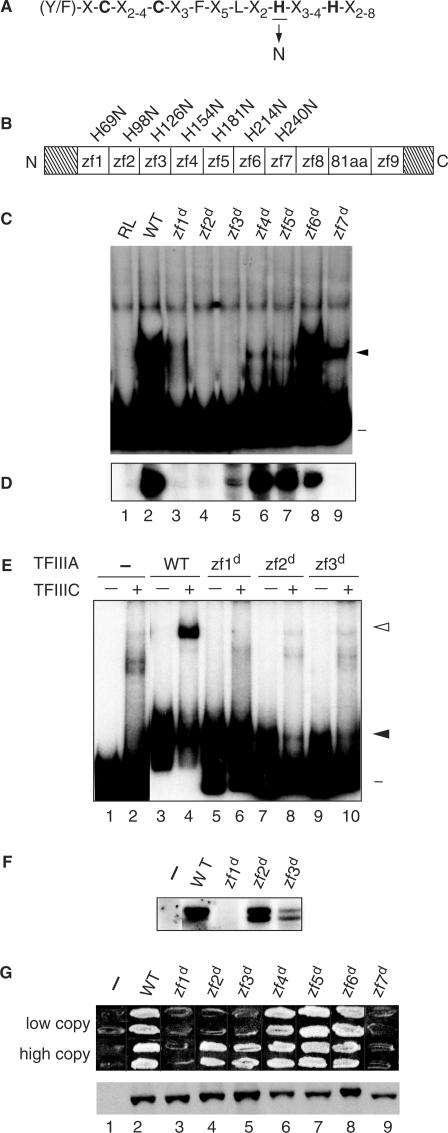
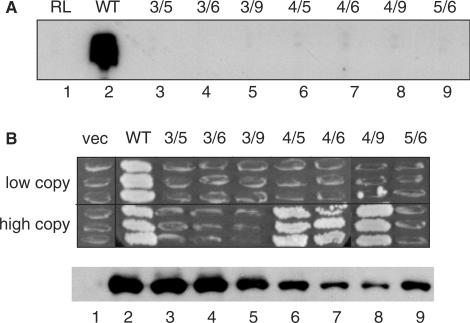
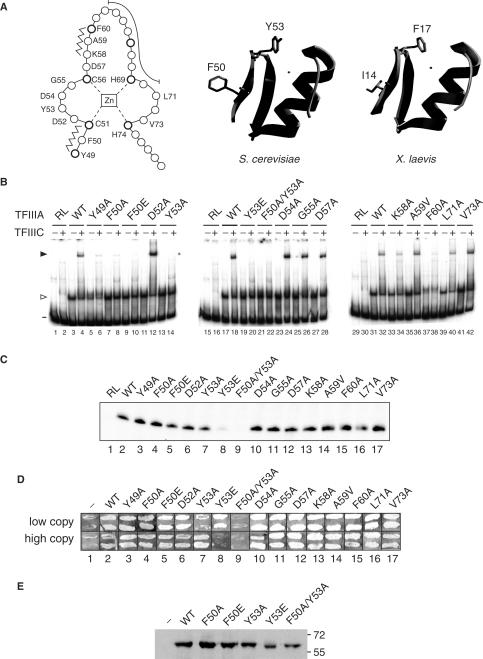
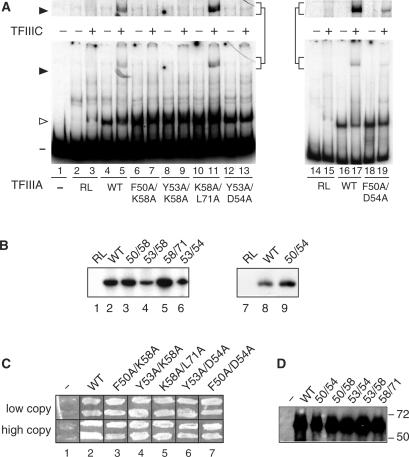
The binding of transcription factor (TF) IIIA to the internal control region of the 5 S RNA gene is the first step in the assembly of a DNA-TFIIIA-TFIIIC- TFIIIB transcription complex, which promotes accurate transcription by RNA polymerase III. With the use of mutations that are predicted to disrupt the folding of a zinc finger, we have examined the roles of zinc fingers 1 through 7 of yeast TFIIIA in the establishment of a functional transcription complex both in vitro and in vivo. Our data indicate that, in addition to their role in DNA binding, the first and seventh zinc fingers contribute other essential roles in the assembly of an active transcription complex. Alanine-scanning mutagenesis identified residues within zinc finger 1 that are not required for DNA binding but are required for incorporation of TFIIIC into the TFIIIA-DNA complex. Although disruption of zinc finger 2 or 3 had a deleterious effect on the activity of TFIIIA both in vitro and in vivo, we found that increasing the level of their in vivo expression allowed these mutant proteins to support cell viability. Disruption of zinc fingers 4, 5 or 6 had minimal effect on the DNA binding and TF activities of TFIIIA.


Figures

Figure 1.
Figure 1.
Zinc finger 1 and zinc finger 7 are essential for the TF activity of TFIIIA. (A) The consensus amino acid sequence of the Cys2His2 zinc finger is given in single-letter code and the zinc-coordinating cysteines and histidines are in bold. The histidine substituted with asparagine to disrupt the structure of individual fingers is underlined. (B) Schematic representation of yeast TFIIIA. The boxes denoted zf1 to zf9 represent the nine zinc fingers; the boxed 81aa denotes the 81-amino-acid domain; and the diagonally stripped boxes represent the 48-amino-acid amino-terminal and the 35-amino-acid carboxyl-terminal regions. The histidine-to-asparagine substitution for each disruption is denoted above the zinc finger. zf = zinc finger. (C) Ability of mutant versions of TFIIIA to bind to the 5 S RNA gene as assessed by EMSA. A radioactively labeled DNA fragment containing the yeast 5 S RNA gene was incubated with in vitro synthesized versions of TFIIIA prior to electrophoresis on a non-denaturing polyacrylamide gel. Lane 1, RL (reticulocyte lysate; in vitro transcription–translation reaction that was not programmed to synthesize TFIIIA); lane 2, WT (wild-type) TFIIIA; lanes 3 through 9, versions of TFIIIA containing a histidine-to-asparagine mutation in the indicated zinc finger. Lane numbers are as given in panel D. The positions of the free DNA (minus sign) and the TFIIIA–DNA complex (closed arrowhead) are indicated on the right. (D) Abilities of mutant versions of TFIIIA to support in vitro transcription of the 5 S RNA gene. In vitro transcription reaction mixtures contained the yeast 5 S RNA gene as template, partially purified yeast TFIIIC, TFIIIB and RNA polymerase III and the version of in vitro synthesized TFIIIA indicated in the corresponding lanes of panel C. The RNAs synthesized in vitro were analyzed on a 7 M urea–10% polyacrylamide gel. The autoradiogram shows the portion of the gel containing 5 S RNA. (E) Abilities of mutant versions of TFIIIA purified from bacteria to bind to the 5 S RNA gene and to recruit TFIIIC to the TFIIIA–DNA complex as assessed by EMSA. A radioactively labeled DNA fragment containing the yeast 5 S RNA gene was incubated with protein extract containing the indicated version of bacterially produced TFIIIA in the absence (odd numbered lanes) or presence (even numbered lanes) of partially purified yeast TFIIIC prior to electrophoresis on a non-denaturing polyacrylamide gel. Lanes are labeled as in panel C with lanes 1 and 2 containing no added bacterial protein. The positions of the free DNA (minus sign), TFIIIA–DNA complexes (solid arrowhead) and TFIIIC–TFIIIA–DNA complexes (arrowhead) are indicated at the right. (F) Abilities of mutant versions of TFIIIA purified from bacteria to support in vitro transcription of the 5 S RNA gene. For details, see legend for Figure 1D. (G) Abilities of mutant versions of TFIIIA to support cell viability. Top panel: a plasmid shuffle assay to test the abilities of mutant versions of TFIIIA to replace wild-type TFIIIA in vivo. Cells of YRW1 that had been transformed with plasmids containing copies of the TFC2 gene were tested for their abilities to grow on medium containing 5-FOA (see Results Section). Each patch contains cells from a different transformant. Two series of plasmids were tested: in the pRS314+ series (labeled ‘low copy’), TFC2 is expressed under the control of its own promoter from a low-copy (CEN ARS) plasmid; in the ΔpG3 series (labeled ‘high copy’), TFC2 is expressed untrol of the strong GPD promoter from a high-copy (2 μ) plasmid. Cells containing the parental vectors not encoding a version of TFIIIA are indicated by the minus sign. Bottom panel: assessment by western blot of in vivo protein levels of mutant versions of TFIIIA. Aliquots of crude lysates prepared from representative YRW1 yeast cells containing the indicated ΔpG3-derived plasmids were separated on a 15% SDS polyacrylamide gel, transferred to a nitrocellulose filter, and probed with anti-TFIIIA antibody. Note that TFIIIA expressed from the pRS314+ series of plasmids is below the level of detection in this blot.
Figure 2.
Figure 2.
Abilities of versions of TFIIIA containing disruptions of two zinc fingers to support TF activity. (A) Abilities of mutant versions of TFIIIA to support in vitro transcription of the 5 S RNA gene. For simplicity, versions of TFIIIA containing histidine-to-asparagine substitutions in multiple fingers are named by the numbers of the disrupted fingers; for example, 3/5 represents TFIIIA with disruptions in zinc fingers 3 and 5 [TFIIIA(H126N/H181N)]. See Figure 1D for details. (B) Abilities of versions of TFIIIA containing disruptions of two zinc fingers to support cell viability. See Figure 1G for details.
Figure 3.
Figure 3.
TF activity of alanine-scanning mutants of the first zinc finger of TFIIIA. (A) A linear representation of the first zinc finger of TFIIIA from S. cerevisiae is on the left. Amino acids are represented by circles, with the position of conserved residues of the zinc finger motif shown in bold. The zinc ion, which is tetrahedrally coordinated by two cysteine and two histidine residues, is boxed in the center of the diagram. Residues that form the short anti-parallel β-sheet of the zinc finger domain are indicated by the zigzag lines, while residues that make up the DNA-binding α-helix are shown by the long curved line. Zinc-coordinating residues and residues that were mutated are identified by their single-letter codes. A ribbon representation of a 3D homology model of the first zinc finger of yeast TFIIIA (residues Y49 to A79) is in the middle. This modeled structure was obtained with the use of the SWISS-MODEL server (63,64) and with the coordinates of the first zinc finger of TFIIIA from X. laevis as the template (17; ExPDB entry 1tf3A; shown on the right). The side chains of F50 and Y53 of the yeast protein and the side chains of the corresponding residues, I14 and F17, of the amphibian protein are shown. The zinc ion, shown by the small circle, is positioned in the yeast structure according to its coordinates in the amphibian protein. (B) Abilities of versions of TFIIIA with alanine-scanning mutations to bind to the 5 S RNA gene and to recruit TFIIIC to the TFIIIA–DNA complex as assessed by EMSA. See Figure 2E for details. The version of TFIIIA used in each reaction is indicated below the gels; RL, reticulocyte transcription–translation reactions not programmed to synthesize TFIIIA; WT, wild-type TFIIIA. The presence (+) or absence (–) of TFIIIC is indicated above the gel. The positions of free DNA (minus sign), TFIIIA–DNA complexes (arrowhead) and TFIIIC–TFIIIA–DNA complexes (solid arrowhead) are shown on the left. (C) Abilities of mutant versions of TFIIIA to support in vitro transcription of the 5 S RNA gene. See Figure 1D for details. (D) Abilities of mutant versions of TFIIIA to support cell viability. See Figure 1G for details. (E) Assessment by western blot analysis of in vivo expression of selected mutant versions of TFIIIA from the ΔpG3 series of plasmids. See Figure 1G for details.
Figure 4.
Figure 4.
Effects of alanine-scanning mutations within the first finger on the activity of TFIIIA. (A) Abilities of versions of TFIIIA with alanine-scanning mutations to bind to the 5 S RNA gene and to recruit TFIIIC to the TFIIIA–DNA complex as assessed by EMSA. See Figure 1E for details. The portion of the gel containing the TFIIIC–TFIIIA–DNA complex is shown at a higher exposure above the full-sized gel. (B) Abilities of mutant versions of TFIIIA to support in vitro transcription of the 5 S RNA gene. See Figure 1D for details. (C) Abilities of mutant versions of TFIIIA to support cell viability. See Figure 1G for details. (D) Assessment by western blot analysis of in vivo expression of mutant versions of TFIIIA from the ΔpG3 series of plasmids. See Figure 1G for details.

References

    1. Geiduschek EP, Kassavetis GA. RNA polymerase III transcription complexes. In: McKnight SL, Yamamoto KR, editors. Transcriptional Regulation. Vol. 1. Cold Spring Harbor, NY: Cold Spring Harbor Laboratory Press; 1992. pp. 247–280.
    1. White RJ. RNA Polymerase III Transcription. Austin: R. G. Landes Company; 1994.
    1. Camier S, Gabrielsen O, Baker R, Sentenac A. A split binding site for transcription factor tau on the tRNA3Glu gene. EMBO J. 1985;4:491–500. - PMC - PubMed
    1. Kassavetis GA, Riggs DL, Negri R, Nguyen LH, Geiduschek EP. Transcription factor IIIB generates extended DNA interactions in RNA polymerase III transcription complexes on tRNA genes. Mol. Cell. Biol. 1989;9:2551–2566. - PMC - PubMed
    1. Stillman DJ, Geiduschek EP. Differential binding of a S. cerevisiae RNA polymerase III transcription factor to two promoter segments of a tRNA gene. EMBO J. 1984;3:847–853. - PMC - PubMed

Publication types

MeSH terms