. 2007 Aug;134(16):2959-68.
doi: 10.1242/dev.006296. Epub 2007 Jul 11.

Regulation of the Arabidopsis root vascular initial population by LONESOME HIGHWAY

Affiliations

Regulation of the Arabidopsis root vascular initial population by LONESOME HIGHWAY

Kyoko Ohashi-Ito et al. Development. 2007 Aug.

Abstract

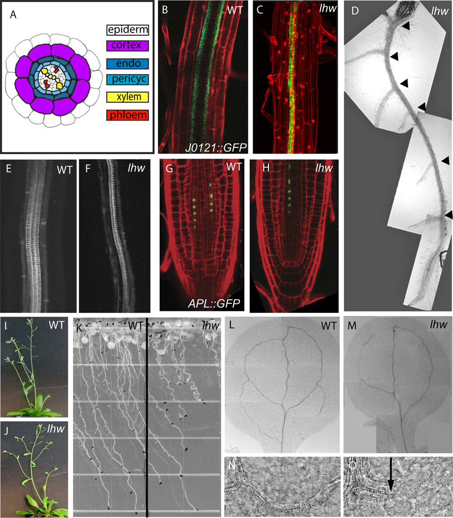
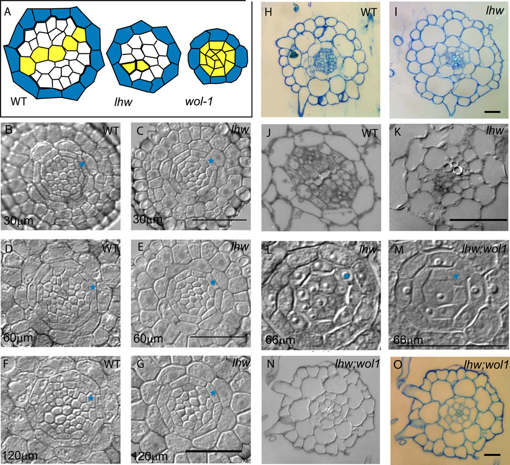
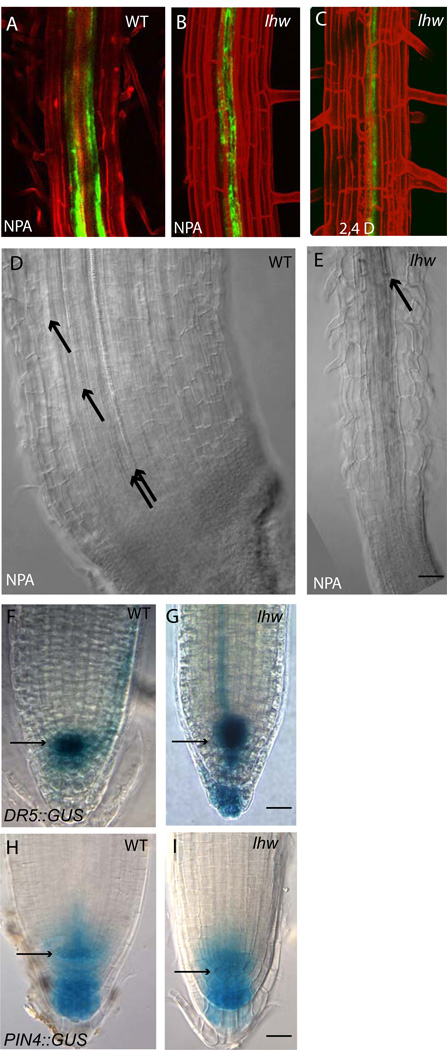
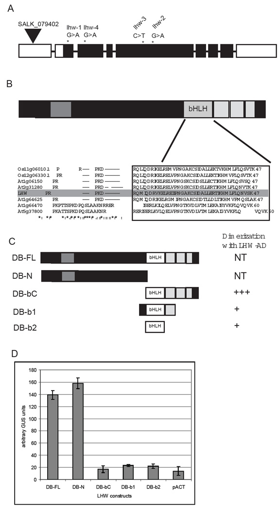
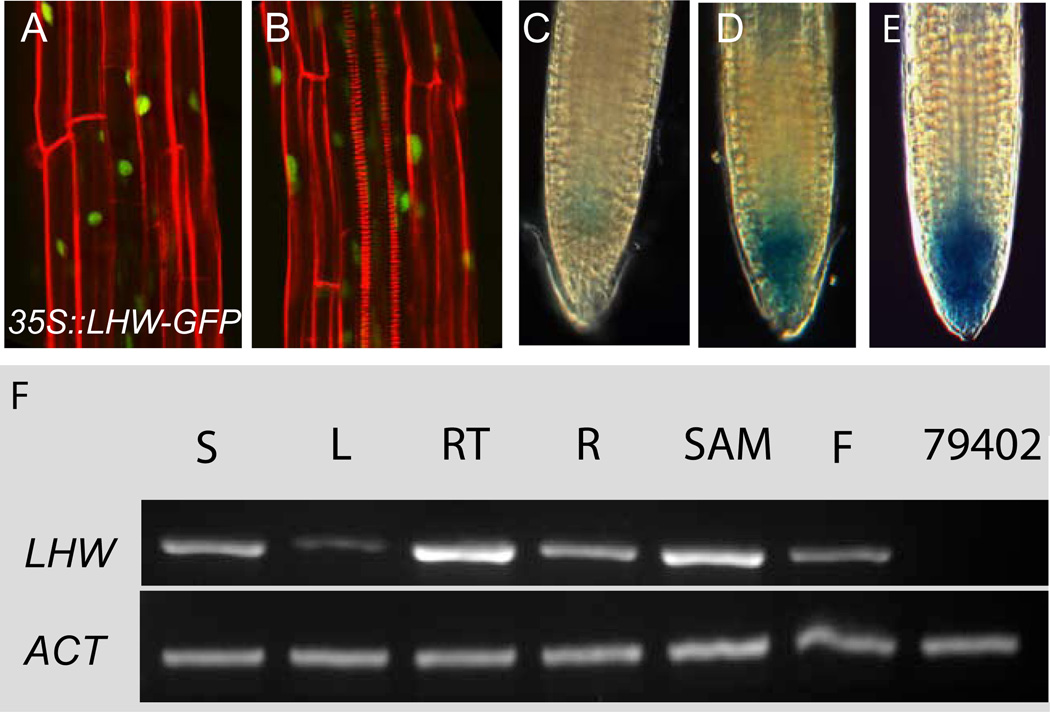
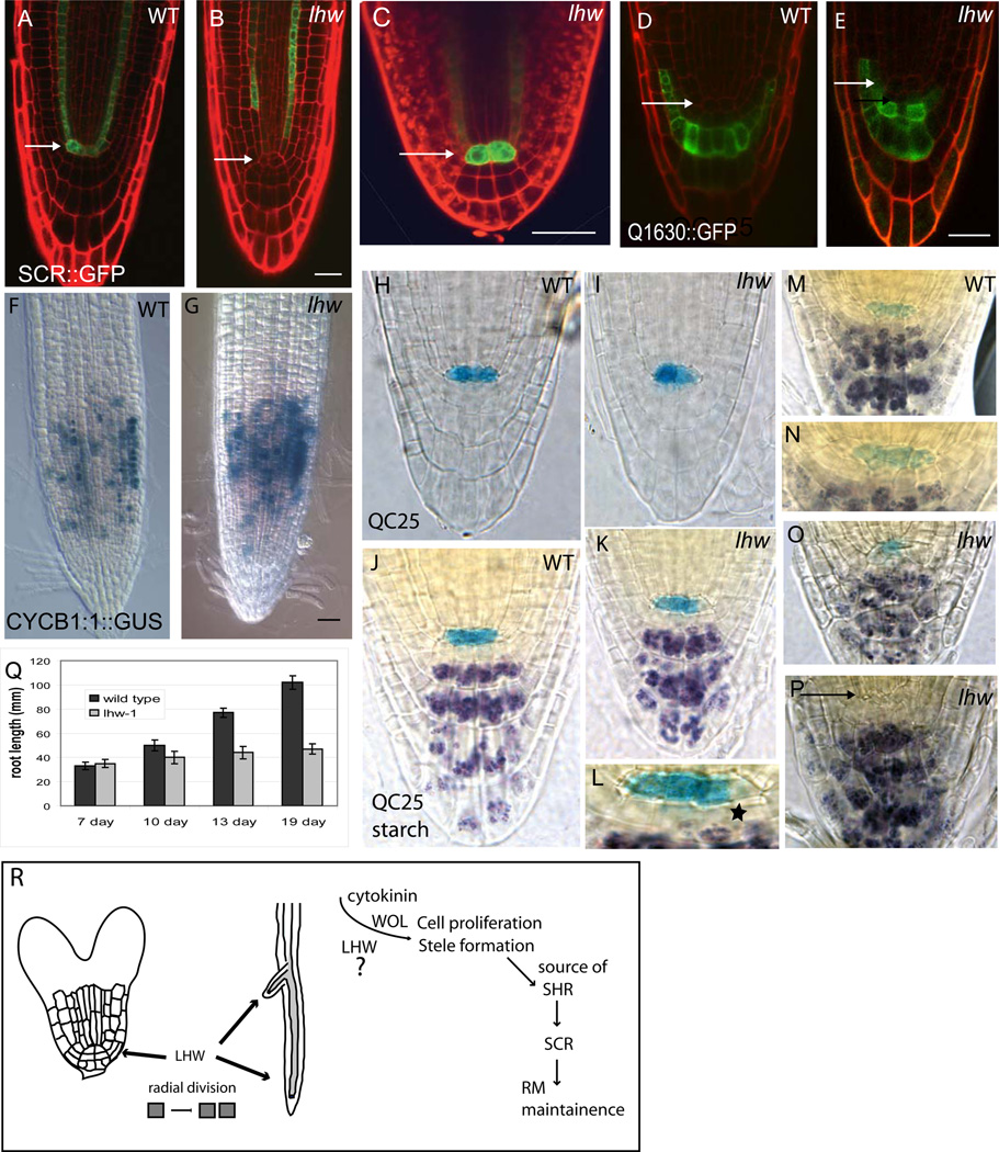
Complex organisms consist of a multitude of cell types arranged in a precise spatial relation to each other. Arabidopsis roots generally exhibit radial tissue organization; however, within a tissue layer, cells are not identical. Specific vascular cell types are arranged in diametrically opposed longitudinal files that maximize the distance between them and create a bilaterally symmetric (diarch) root. Mutations in the LONESOME HIGHWAY (LHW) gene eliminate bilateral symmetry and reduce the number of cells in the center of the root, resulting in roots with only single xylem and phloem poles. LHW does not appear to be required for the creation of any specific cell type, but coordinately controls the number of all vascular cell types by regulating the size of the pool of cells from which they arise. We cloned LHW and found that it encodes a protein with weak sequence similarity to basic helix-loop-helix (bHLH)-domain proteins. LHW is a transcriptional activator in vitro. In plants, LHW is nuclear-localized and is expressed in the root meristems, where we hypothesize it acts independently of other known root-patterning genes to promote the production of stele cells, but might also indirectly feed into established regulatory networks for the maintenance of the root meristem.


Figures

Figure 1
Figure 1. Phenotype of LONESOME HIGHWAY mutants
(A) Cross section diagram of a mature Arabidopsis root; tissues are arranged radially from outside in: epidermis (white), cortex (light purple), endodermis (dark blue) and stele. The stele consists of a ring of pericycle cells (light blue), surrounding xylem (yellow) and phloem (red) arranged in bilateral symmetry. (B–C) Confocal images of wild type (B) and lhw-1 (C) expressing xylem-associated pericycle marker J0121::GFP in green. Roots are counterstained with propidum iodide (PI, red) to visualize outlines of cells. (D) Brightfield image of lhw-1 root with all lateral roots (black arrowheads) emerging from single side of primary root. (E–F) Confocal image of basic fucshin staining of xylem in wildtype (E) and lhw-1 (F). (G–H) Confocal images of wild type (G) and lhw-1 (H) expressing phloem marker APLpro::APL-GFP in green. (I–J) Whole plant phenotypes of WT Col (I) and lhw SALK_079402 (J). (K) Root growth of wild type (left) and lhw-1 (right) on agar plates at 20-dpg. Note root waving and short root phenotypes in lhw-1. (L–M) vascular pattern in mature (13 dpg) WT (L) and lhw-1 (M) cotyledons (N–O) higher magnification images of xylem from images L and M, respectively. Black arrow points to end of mature xylem elements, to the right, elongated cells typical of procambium are still seen. For each marker, the WT and lhw image pair are at same magnification.
Figure 2
Figure 2. Cross sections of lhw roots and hypocotyls
(A) Schematic of stele in cross sections from WT, lhw and wol-1; pericycle in blue and xylem in yellow; WT and lhw were traced from 2F and 2G, respectively. (B–K) Brightfield images of WT and lhw root sections taken at increasing distance from tip of root; one pericycle cell marked with blue star for orientation. (B) WT, 30 µm (C) lhw-2, 30 µm (D) WT, 60 µm (E) lhw-2, 60 µm (F) WT, 120 µm (G) lhw-2, 120 µm. (H–I) Toluidine blue staining of WT (H) and lhw-2 (I) in mature zone. (J–K) section through lower third of hypocotyl in WT (J) and lhw-2 (K). (L) Center of wol-1 root, 66 µm from tip (M) Center of wol-1;lhw-1 root, 66 µm from tip. (N–O) DIC and toluidine blue images of wol-1;lhw-1 mature roots showing the very reduced stele filled with xylem elements. Each image pair is at the same magnification. Scale bars = 20 µm.
Figure 3
Figure 3. Measures of auxin response in lhw
(A–C) confocal images of WT (A) and lhw-1 (B–C) expressing J0121::GFP. (A–B) treatment with 20 µM NPA for 7 days (C) treatment with 30nM 2,4D for seven days. (D–E) DIC images of roots treated with 20 µM NPA for 21 days. Black arrows point to xylem elements. (F–I) Brightfield images 7-dpg seedling root tips; (F-G) DR5::GUS expression; (H–I) PIN4::GUS expression. Each image pair (and A–C) is at the same magnification. Scale bars = 20 µm.
Figure 4
Figure 4. LHW gene and protein structure and behavior in two hybrid screen
(A) LHW gene structure with exons represented as boxes and introns as lines. LHW coding region is in black. The location and nature of lhw mutant alleles is indicated above the exons. (B) LHW protein structure. Two domains conserved with other plant proteins are indicated as grey boxes. Part of the C-terminal conserved region resembles the bHLH domain of transcriptional regulators. A sequence alignment of the putative bHLH domain (boxed) is diagrammed for LHW and related proteins from Arabidopsis and rice. (C) Graphical representation of LHW protein fragments used in yeast two hybrid assay and their ability to dimerize with full length LHW. (D) GAL4 transcriptional activation activity of LHW variants; pACT is included as a negative control. GUS measurements based on 4 replicates/sample. Error bars ± SEM.
Figure 5
Figure 5. LHW expression
(A–B) Confocal images of lhw-2 roots expressing 35S::LHW-GFP in nuclei (green). Expression of this transgene is sufficient to rescue the lhw-2 xylem phenotype (B). (C-E) LHWpro::GUS reporter expression in root tips, staining for 1hr, 4hr and 8hr, respectively (F). RT-PCR of LHW and actin (ACT) expression. S, 14-dpg seedling; L, expanded rosette leaf; RT, 5mm of 14-dpg root tip; R, whole 14-dpg root; SAM, transition SAM and youngest leaves; F, flower stage 8-15; 79402, 14-dpg seedling of SALK_79402.
Figure 6
Figure 6. LHW effects on RM meristem establishment and maintenance
(A–I) Meristem markers in WT and lhw-1 7-dpg seedlings (A–B) SCR::GFP expression (C) SCR::GFP in torpedo stage lhw-1 embryo (D–E) Columella differentiation marker Q1630::GFP. (F–G) CYCB1;2::GUS expression (H–P) QC25::GUS (blue) and starch granules (purple) mark degeneration of lhw meristem over time (H–I; 7dpg, J–L; 13 dpg; M–P; 18 dpg). L is higher magnification image of K; start indicates starch granule containing cells adjacent to QC25 marked cells. N is higher magnification of M. (Q) Graph of WT (dark grey) and lhw-1 (light grey) root growth over time. For each marker, the WT and lhw image pair are the same magnification. Arrows point to QC cells. (R) Model for LHW action in generating vascular pattern. LHW is required to establish the radial extent of the root vascular tissues in the embryo and promotes postembryonic divisions in these tissues. LHW therefore acts as a meristem size-control protein for the center of the root. LHW and WOL are both required for these cell divisions, but appear to act at least somewhat independently. We propose that the eventual slowing down of longitudinal growth in lhw mutant roots is not due to direct requirement for LHW in meristem maintenance, but because LHW is required to create the tissue that normally produce SHR. Without adequate levels of SHR, SCR is not maintained in the QC and meristems eventually terminate. Each image pair (A-K and M, O, P) at the same magnification. Scale bars = 30 µm. Error bars in Q ± SEM

References

    1. Aida M, Beis D, Heidstra R, Willemsen V, Blilou I, Galinha C, Nussaume L, Noh YS, Amasino R, Scheres B. The PLETHORA Genes Mediate Patterning of the Arabidopsis Root Stem Cell Niche. Cell. 2004;119:109–120. - PubMed
    1. Birnbaum K, Jung JW, Wang JY, Lambert GM, Hirst JA, Galbraith DW, Benfey PN. Cell Type-Specific Expression Profiling in Plants Via Cell Sorting of Protoplasts from Fluorescent Reporter Lines. Nat. Methods. 2005;2:615–619. - PubMed
    1. Birnbaum K, Shasha DE, Wang JY, Jung JW, Lambert GM, Galbraith DW, Benfey PN. A Gene Expression Map of the Arabidopsis Root. Science. 2003;302:1956–1960. - PubMed
    1. Blilou I, Frugier F, Folmer S, Serralbo O, Willemsen V, Wolkenfelt H, Eloy NB, Ferreira PC, Weisbeek P, Scheres B. The Arabidopsis HOBBIT Gene Encodes a CDC27 Homolog that Links the Plant Cell Cycle to Progression of Cell Differentiation. Genes Dev. 2002;16:2566–2575. - PMC - PubMed
    1. Blilou I, Xu J, Wildwater M, Willemsen V, Paponov I, Friml J, Heidstra R, Aida M, Palme K, Scheres B. The PIN Auxin Efflux Facilitator Network Controls Growth and Patterning in Arabidopsis Roots. Nature. 2005;433:39–44. - PubMed

Publication types

MeSH terms