. 2007 Sep;81(18):9790-800.
doi: 10.1128/JVI.00530-07. Epub 2007 Jul 11.

Differential type I interferon induction by respiratory syncytial virus and influenza a virus in vivo

Affiliations

Differential type I interferon induction by respiratory syncytial virus and influenza a virus in vivo

Nancy A Jewell et al. J Virol. 2007 Sep.

Abstract

Type I interferon (IFN) induction is an immediate response to virus infection, and very high levels of these cytokines are produced when the Toll-like receptors (TLRs) expressed at high levels by plasmacytoid dendritic cells (pDCs) are triggered by viral nucleic acids. Unlike many RNA viruses, respiratory syncytial virus (RSV) does not appear to activate pDCs through their TLRs and it is not clear how this difference affects IFN-alpha/beta induction in vivo. In this study, we investigated type I IFN production triggered by RSV or influenza A virus infection of BALB/c mice and found that while both viruses induced IFN-alpha/beta production by pDCs in vitro, only influenza virus infection could stimulate type I IFN synthesis by pDCs in vivo. In situ hybridization studies demonstrated that the infected respiratory epithelium was a major source of IFN-alpha/beta in response to either infection, but in pDC-depleted animals only type I IFN induction by influenza virus was impaired.


Figures
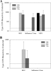
FIG. 1.
FIG. 1.
Type I IFN induction in lung epithelial cell lines. (A) Levels of type I IFN bioactivity measured in supernatants from a human epithelial cell line, A549, infected with RSV, influenza virus, or NDV. Samples were taken at 24, 48, and 72 h after infection. NDV was included as a positive control. (B) Levels of type I IFN bioactivity in supernatants from a murine epithelial cell line, LA-4, infected with RSV and influenza virus. Samples were taken at 24, 48, and 72 h after infection. The limit of detection of IFN activity in the cells is indicated by the dashed line. Error bars are based upon duplicate experiments.
FIG. 2.
FIG. 2.
Infection with influenza virus induced prolonged type I IFN production. (A) Mice were inoculated i.n. with 106 PFU of either RSV or WSN virus. Lungs were harvested 24, 48, or 72 h postinfection, and lung homogenates were assayed for antiviral activity against vesicular stomatitis virus. Samples from WSN virus- and RSV-infected animals are shown as white and black bars, respectively. The limit of detection of IFN activity in the lung homogenates is indicated by the dashed line. (B) Levels of IFN-β protein were determined by ELISA from BAL fluid samples in a repeat experiment. (C) The dose dependence of type I IFN bioactivity induced by WSN virus infection was determined by bioassay of BAL fluid samples. Error bars are based upon a sample size of three mice at each time point for each virus tested.
FIG. 3.
FIG. 3.
Viral antigen loads in lung tissues at 24 and 96 h. Mice were infected i.n. with either 107 PFU of RSV (A and C) or 104 PFU of WSN virus (B and D). Representative lung sections from tissue harvested at 24 and 96 h postinfection are pictured, both H&E stained and immunostained. At 24 h (A and B), rare antigen-positive cells are present focally within bronchiolar epithelium following infection with either virus. By 96 h (C and D) postinfection with either pathogen, diffuse immunostaining is present. In RSV-infected mice (C), inflammation is sparse and infection is seen primarily in the cells lining the alveolar spaces. In WSN virus-infected animals (D), inflammatory cells surround bronchioles and vessels and there is intense immunostaining of both bronchiolar and alveolar epithelia. In addition, a large amount of antigen-positive cell debris is present in the alveolar spaces.
FIG. 4.
FIG. 4.
Type I IFN induced by RSV infection declines after 24 h. Cohorts of mice (n = 5) were infected with 107 PFU of RSV or 104 PFU of WSN virus or mock infected with uninfected HEp2 cell lysate. BAL fluid samples from infected animals were harvested at 24, 48, or 72 h and assayed for IFN-α (A) and IFN-β (B) protein concentrations by ELISA.
FIG. 5.
FIG. 5.
Type I IFN induction requires virus replication. Mice were infected with equivalent doses of live and UV-inactivated virus (RSV, 107 PFU; influenza virus, 105 PFU), and lungs were harvested at 24, 48, 72, and 96 h postinfection. IFN activity was measured at 24 h (light gray bars), 48 h (white bars), 72 h (black bars), and 96 h (dark gray bars) postinfection. Error bars are based upon a sample size of five mice at each time point for each virus tested.
FIG. 6.
FIG. 6.
No inflammation is triggered by i.n. instillation of UV-inactivated virus. Following instillation of live and UV-inactivated virus preparations, mice were monitored by histology for the presence of lung inflammation, epithelial cytopathology, or evidence of viral antigen. No evidence of infection was found in these mice or mock-infected controls. The left panel shows an H&E-stained section from a lung harvested 96 h following inoculation with UV-inactivated RSV. The right panel shows a serial section stained for the presence of RSV antigens. No viral proteins were detected by immunohistochemistry.
FIG. 7.
FIG. 7.
RSV and influenza virus have distinct type I IFN induction profiles. Mice were infected i.n. with either 107 PFU of RSV (A) or 104 PFU of WSN virus (B), and their lungs were harvested at 24, 48, 72, or 96 h postinfection. Lung homogenates were assayed for type I IFN activity and for viral titers. Levels of type I IFN activity are plotted on the left y axis and are indicated by squares. Viral titers are plotted on the right y axis and are indicated by circles. Error bars are based upon a sample size of five mice at each time point. Black squares and circles, RSV (A). White squares and circles, WSN virus (B).
FIG. 8.
FIG. 8.
Infected lung epithelial cells are a major source of type I IFNs. Mice were i.n. infected with either 107 PFU/ml RSV or 104 PFU/ml influenza virus, and tissues were harvested at 48 h postinfection. (A) In situ hybridization with a DIG-labeled murine IFN-α4 riboprobe was used to detect the presence of type I IFN transcripts in the lung tissue and adjacent lymph nodes. (B) Accumulation of the various DC subsets at 24-h intervals following i.n. infection of mice with 104 PFU of influenza A virus. CD11c = total DCs; CD11c, CD11b = myeloid DCs; CD11c, CD8 = antigen-presenting DCs; CD11c, B220 = pDCs. Error bars are based upon a sample size of five mice at each time point for each dose of virus tested.
FIG. 9.
FIG. 9.
Reduced IFN-α/β production by RSV-treated DCs in vitro. BMDCs cultured with FLT-3 ligand were mock infected or infected with either RSV (live or UV inactivated) or WSN virus (live or UV inactivated) at an MOI of 1. At 24 h postinfection, infected-DC supernatants were assayed for IFN-α and IFN-β protein concentrations by ELISA.
FIG. 10.
FIG. 10.
pDC depletion does not alter type I IFN synthesis in RSV-infected mice. Mice were infected with virus (106 PFU of WSN virus or 107 PFU of RSV) following either pDC depletion by i.p. injection of monoclonal antibody PDCA-1 or i.p. injection of rat IgG, which was used as a control for antibody treatment. (Α) Extent of pDC depletion was monitored by fluorescence-activated cell sorter analysis of blood taken from individual mice (n = 4 animals per group). (B) Type I IFN levels in lung homogenates were determined by ELISA 24 h after infection (n = 4 mice per group). Both IFN-α (P = 0.0019) and IFN-β (P = 0.003) were significantly lower in pDC-depleted, WSN virus-infected animals, but there were no significant differences in lung samples from RSV-infected mice with or without PDCA-1 treatment. (C) Lung IFN-α/β levels were measured at multiple time points following infection of IFNAR−/− mice with RSV (107 PFU) or WSN virus (104 PFU) (n = 5 animals per group).

References

    1. Asselin-Paturel, C., A. Boonstra, M. Dalod, I. Durand, N. Yessaad, C. Dezutter-Dambuyant, A. Vicari, A. O'Garra, C. Biron, F. Brière, and G. Trinchieri. 2001. Mouse type I IFN-producing cells are immature APCs with plasmacytoid morphology. Nat. Immunol. 2:1144-1150. - PubMed
    1. Asselin-Paturel, C., G. Brizard, K. Chemin, A. Boonstra, A. O'Garra, A. Vicari, and G. Trinchieri. 2005. Type I interferon dependence of plasmacytoid dendritic cell activation and migration. J. Exp. Med. 201:1157-1167. - PMC - PubMed
    1. Baccam, P., C. Beauchemin, C. Macken, F. Hayden, and A. Perelson. 2006. Kinetics of influenza A virus infection in humans. J. Virol. 80:7590-7599. - PMC - PubMed
    1. Barchet, W., M. Cella, C. Odermatt, C. Asselin-Paturel, M. Colonna, and U. Kalinke. 2002. Virus-induced interferon alpha production by a dendritic cell subset in the absence of feedback signaling in vivo. J. Exp. Med. 195:507-516. - PMC - PubMed
    1. Biron, C. A., K. B. Nguyen, G. C. Pien, L. P. Cousens, and T. P. Salazar-Mather. 1999. Natural killer cells in antiviral defense: function and regulation by innate cytokines. Annu. Rev. Immunol. 17:189-220. - PubMed

Publication types

MeSH terms