. 2007 Sep;81(18):9900-10.
doi: 10.1128/JVI.00909-07. Epub 2007 Jul 11.

Mutations in the stalk of the measles virus hemagglutinin protein decrease fusion but do not interfere with virus-specific interaction with the homologous fusion protein

Affiliations

Mutations in the stalk of the measles virus hemagglutinin protein decrease fusion but do not interfere with virus-specific interaction with the homologous fusion protein

Elizabeth A Corey et al. J Virol. 2007 Sep.

Abstract

The hemagglutinin (H) protein of measles virus (MV) mediates attachment to cellular receptors. The ectodomain of the H spike is thought to consist of a membrane-proximal stalk and terminal globular head, in which resides the receptor-binding activity. Like other paramyxovirus attachment proteins, MV H also plays a role in fusion promotion, which is mediated through an interaction with the viral fusion (F) protein. The stalk of the hemagglutinin-neuraminidase (HN) protein of several paramyxoviruses determines specificity for the homologous F protein. In addition, mutations in a conserved domain in the Newcastle disease virus (NDV) HN stalk result in a sharp decrease in fusion and an impaired ability to interact with NDV F in a cell surface coimmunoprecipitation (co-IP) assay. The region of MV H that determines specificity for the F protein has not been identified. Here, we have adapted the co-IP assay to detect the MV H-F complex at the surface of transfected HeLa cells. We have also identified mutations in a domain in the MV H stalk, similar to the one in the NDV HN stalk, that also drastically reduce fusion yet do not block complex formation with MV F. These results indicate that this domain in the MV H stalk is required for fusion but suggest either that mutation of it indirectly affects the H-dependent activation of F or that the MV H-F interaction is mediated by more than one domain in H. This points to an apparent difference in the way the MV and NDV glycoproteins interact to regulate fusion.


Figures

FIG. 1.
FIG. 1.
Diagram of MV H structure, including the amino acid sequences of the HR-like region (residues 84 to 105) and region 244 to 250. Individual mutations introduced into these regions are listed beneath the wt sequence. tm, transmembrane; C139 and C154, cysteines 139 and 154 involved in intermonomer disulfide bonds.
FIG. 2.
FIG. 2.
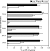
Cell surface expression (CSE) and functional characteristics of the MV H proteins with alanine substitutions in region 244 to 250. CSE was quantitated by flow cytometry with MAbs specific for antigenic sites in the ectodomain of the protein. HAd activity was quantitated by the ability of the transfected HeLa cells to adsorb AGM erythrocytes. The abilities of the mutated H proteins to complement MV Fcsm in the promotion of membrane fusion were determined by a content-mixing assay. For each assay, the background detected in cells transfected with vector has been subtracted. All data points represent the means of at least three independent experiments and are expressed relative to the activities of the wt proteins.
FIG. 3.
FIG. 3.
Comparison of the amino acid sequences in the HR-like domains in the stalks of various paramyxovirus attachment glycoproteins. The numbers (1 to 4) above the sequences indicate the heptadic residues, which are also in bold print. The highly conserved proline and leucine residues are indicated in large print. The sequences and relevant references are as follows: MV (1), CDV (21), NDV (26), hPIV3 (12), and mumps virus (MuV) (46).
FIG. 4.
FIG. 4.
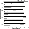
Cell surface expression (CSE) and functional characteristics of the MV H proteins with mutations in the heptadic residues of the HR-like domain. Assays were performed as described in the legend to Fig. 2. All data points represent the means of at least three independent experiments and are expressed relative to the activities of the wt proteins.
FIG. 5.
FIG. 5.
Cell surface expression (CSE) and functional characteristics of the MV H proteins with mutations at P94 and L95 in the HR-like domain. Assays were performed as described in the legend to Fig. 2. All data points represent the means of at least three independent experiments and are expressed relative to the activities of the wt proteins.
FIG. 6.
FIG. 6.
Cell surface expression (CSE) and functional characteristics of the MV H proteins with mutations within the IRs of the HR-like domain. Assays were performed as described in the legend to Fig. 2. All data points represent the means of at least three independent experiments and are expressed relative to the activities of the wt proteins.
FIG. 7.
FIG. 7.
Correlation of ability to promote fusion with HAd activity of MV H proteins with mutations in the HR-like domain in the stalk. Based on this plot, the mutated proteins can be divided into three groups: (1) no significant deficiencies in receptor binding or fusion promotion, (2) significant deficiencies in both activities, and (3) significant deficiencies in fusion promotion but not receptor binding.
FIG. 8.
FIG. 8.
Detection of formation of a complex of wt MV H and Fcsm at the surface of HeLa cells by using a co-IP assay. The surface proteins of transfected cells were biotinylated, and then the cells were lysed with lauryl maltoside. The lysates were split into two equal aliquots and subjected to the co-IP protocol by using a mixture of two MAbs against the H protein and a polyclonal antiserum against the cytoplasmic tail of F (the first lane in each pair) or the F antiserum alone (the second lane in each pair). Immunoprecipitates were displayed on reducing SDS-polyacrylamide gels.
FIG. 9.
FIG. 9.
Detection of formation of complexes between MV H mutants with MV Fcsm at the cell surface. (A) Co-IP of MV H proteins carrying mutations in the heptadic residues with MV F. (B) Co-IP of MV H proteins carrying mutations at P94 and L95 with MV F.
FIG. 10.
FIG. 10.
Co-IP of MV H proteins carrying mutations in the IR with MV F. (A) Co-IP of L92A- and T93A-mutated proteins. (B) Co-IP of proteins carrying a mutation at position 96, 97, or 99. The experiments were performed as described in the legend to Fig. 8.
FIG. 11.
FIG. 11.
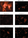
Promotion of lipid mixing by MV H proteins with mutations in the HR-like domain. The extent of lipid mixing is shown by the spread of the dye from R18-labeled AGM erythrocytes into the cell membranes of transfected HeLa cells. Labeled erythrocytes were added to each monolayer, and then the cells were incubated on ice for 30 min. Fusion between the transfected cells and erythrocytes was initiated by transferring the cells to 37°C. After 30 min, the cells were washed, and then images were acquired immediately with a 20× objective by using fluorescent microscopy.

References

    1. Alkhatib, G., and D. J. Briedis. 1986. The predicted primary structure of the measles virus hemagglutinin. Virology 150:479-490. - PubMed
    1. Bagai, S., and R. A. Lamb. 1995. Quantitative measurement of paramyxovirus fusion: differences in requirements of glycoproteins between simian virus 5 and human parainfluenza virus 3 or Newcastle disease virus. J. Virol. 69:6712-6719. - PMC - PubMed
    1. Bossart, K. N., L. F. Wang, M. N. Flora, K. B. Chua, S. K. Lam, B. T. Eaton, and C. C. Broder. 2002. Membrane fusion tropism and heterotypic functional activities of the Nipah virus and Hendra virus envelope glycoproteins. J. Virol. 76:11186-11198. - PMC - PubMed
    1. Cathomen, T., H. Y. Naim, and R. Cattaneo. 1998. Measles viruses with altered envelope protein cytoplasmic tails gain cell fusion competence. J. Virol. 72:1224-1234. - PMC - PubMed
    1. Cattaneo, R., and J. K. Rose. 1993. Cell fusion by the envelope glycoproteins of persistent measles viruses which caused lethal human brain disease. J. Virol. 67:1493-1502. - PMC - PubMed

Publication types

MeSH terms

Substances