. 2007 Sep;81(18):9778-89.
doi: 10.1128/JVI.00360-07. Epub 2007 Jul 11.

Type I interferon inhibition and dendritic cell activation during gammaherpesvirus respiratory infection

Affiliations

Type I interferon inhibition and dendritic cell activation during gammaherpesvirus respiratory infection

Janet L Weslow-Schmidt et al. J Virol. 2007 Sep.

Abstract

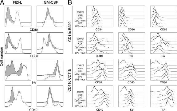
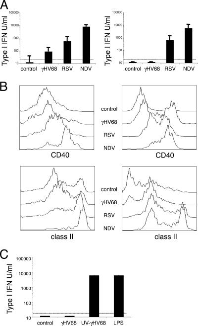
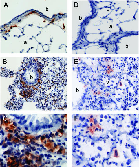
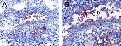
The respiratory tract is a major mucosal site for microorganism entry into the body, and type I interferon (IFN) and dendritic cells constitute a first line of defense against viral infections. We have analyzed the interaction between a model DNA virus, plasmacytoid dendritic cells, and type I IFN during lung infection of mice. Our data show that murine gammaherpesvirus 68 (gammaHV68) inhibits type I IFN secretion by dendritic cells and that plasmacytoid dendritic cells are necessary for conventional dendritic cell maturation in response to gammaHV68. Following gammaHV68 intranasal inoculation, the local and systemic IFN-alpha/beta response is below detectable levels, and plasmacytoid dendritic cells are activated and recruited into the lung with a tissue distribution that differs from that of conventional dendritic cells. Our results suggest that plasmacytoid dendritic cells and type I IFN have important but independent roles during the early response to a respiratory gammaHV68 infection. gammaHV68 infection inhibits type I IFN production by dendritic cells and is a poor inducer of IFN-alpha/beta in vivo, which may serve as an immune evasion strategy.


Figures

FIG. 1.
FIG. 1.
Dendritic cell activation in response to γHV68 infection. (A) Bone marrow-derived dendritic cells grown in the presence of Flt3-L (left column) or GM-CSF (right column) were infected (empty histograms) or not infected (gray histograms) with γHV68 as described in Materials and Methods. Forty hours later, the cells were surface stained with antibodies against CD11c and the indicated activation markers to analyze their fluorescence intensity on a flow cytometer. (B) Bone marrow-derived dendritic cells grown in the presence of Flt3-L were infected or not infected with γHV68 and stimulated with CpG-ODN or LPS as described in Materials and Methods. Forty-eight hours after treatment, the cells were harvested and stained with antibodies against the indicated cell surface markers to analyze their fluorescence intensity. Dendritic cells were previously gated as plasmacytoid dendritic cells (CD11c+ B220+) or conventional dendritic cells (CD11c+ CD11b+). The data are representative of three independent experiments. The data shown in A and B are from two independent experiments.
FIG. 2.
FIG. 2.
γHV68 inhibits type I IFN production by dendritic cells. (A) Type I IFN induction by γHV68, RSV, or NDV was measured in cell culture supernatants 40 h after infection using an IFN-α/β bioassay. The dendritic cells were grown in the presence of GM-CSF (left column) or Flt3-L (right column). (B) The relative abilities of γHV68, RSV, or NDV to induce maturation of dendritic cells were tested using bone marrow-derived dendritic cells grown in the presence of GM-CSF (left column) or Flt3-L (right column). The cell cultures were infected as described in Materials and Methods, and 40 h later, the cells were surface stained with antibodies against CD11c, CD40, and I-A. The histograms shown have been previously gated as CD11c+ cells. (C) Type I IFN induction by UV-inactivated γHV68 in dendritic cell cultures supplemented with GM-CSF. The data presented are the means and standard deviations of triplicate dendritic cell cultures.
FIG. 3.
FIG. 3.
Lung dendritic cell location after γHV68 infection. Lungs were sampled from naïve mice (A and D) or from γHV68-infected mice at 7 days postinfection (B and E). Serial sections were stained with anti-CD11c (left panels) or anti-mPDCA-1 (right panels) as described in Materials and Methods. Objective magnification, ×20. C and F show a detail of the tissue area adjacent to the bronchi (b) or blood vessel (v) from B and E, respectively. One representative staining out of three mice per group is shown from three independent experiments. a, arteriole.
FIG. 4.
FIG. 4.
Location of viral antigens in the lung of γHV68-infected mice. Lungs were sampled from γHV68-infected mice at 7 days postinfection, and sections were stained with anti-γHV68 M3 antiserum. (A) Area of inflammation in the lung parenchyma. Magnification, ×20. (B) Detail of the bronchi (b) from A. Magnification, ×40.
FIG. 5.
FIG. 5.
Identification of dendritic cell subsets in respiratory tract mPDCA-1+ cells. (A) Expression of CD8α and CD11b in previously gated CD11c+ cells defines different dendritic cell subsets in the lung, bone marrow, and spleen of mice 5 days after γHV68 infection: 1, plasmacytoid dendritic cells; 2, CD8 conventional dendritic cells; 3, conventional CD11b dendritic cells. (B) B220 cell surface expression on mouse dendritic cell subsets as previously gated in A. Data are representative of three independent experiments.
FIG. 6.
FIG. 6.
Dendritic cell migration into the lung and spleen in response to γHV68 infection. (A) Time course analysis of the absolute dendritic cell numbers in bone marrow, lung, and spleen after γHV68 infection. The data presented are the means and standard deviations of three independent experiments, each containing three mice. (B) Plasmacytoid dendritic cells migrate into the lung and spleen after γHV68 infection. Numbers indicate the percentages of cells inside the gate. One representative of three experiments is shown. (C) Time course analysis of the frequency of plasmacytoid dendritic cells (DC) in bone marrow, lung, and spleen after γHV68 infection. The data presented are the means and standard deviations of three independent experiments, each containing three mice. In all the panels shown, dendritic cells were analyzed as CD11c+ and lineage-negative (CD3, CD19, and NK1.1) nonautofluorescent cells.
FIG. 7.
FIG. 7.
Dendritic cell maturation and IFN-α/β production in γHV68-infected mice. (A) Dendritic cell subsets undergo differential maturation in the lung in response to γHV68 infection. Lung dendritic cells were analyzed at different time points after γHV68 infection (days 0 to 7) for the level of cell surface expression of several activation markers (CD80, CD86, CD40, and I-A). Histograms are previously gated as CD11c+ B220+ (plasmacytoid dendritic cells) (first row), CD11c+ CD11b+ (conventional dendritic cells) (second row), or CD11c+ CD8a+ (conventional dendritic cells) (third row). (B) Type I IFN bioactivity in BAL fluid of γHV68- and NDV-infected mice at different time points after infection. (C) Type I IFN bioactivity in serum of γHV68- and NDV-infected mice at different time points after infection. The data presented are the means and standard deviations for three to four mice.
FIG. 8.
FIG. 8.
Role of type I IFN signaling in virus control and dendritic cell recruitment and activation in the lung. (A) Infectious virus titers in the lung of 129SvEv or IFN-α/βR−/− mice were determined by plaque assay on day 5 after virus inoculation. (B) Frequency of CD11c+ dendritic cells in the lung of γHV68-infected mice. (C) Representative histograms of the level of expression of class II molecules on CD11c+ cells in the lung. Numbers indicate the percentage of cells inside the gate. (D) Frequency of class II expression on CD11c+ cells in the lung. FACS analysis was performed on naïve and γHV68-infected mice on day 7. The data presented are the means and standard deviations for three individual mice.
FIG. 9.
FIG. 9.
Absence of type I IFN-producing cells in lung and draining lymph nodes of γHV68-infected mice. Lung (A to F) and MLN (G to J) tissue sections from influenza virus-infected (A to C, G, and H) or γHV68-infected (D to F, I, and J) mice (days 2, 3, and 7) or control mice were labeled with riboprobes of murine IFN-α4 and IFN-β genes. (A) Influenza virus-infected lung, day 2, with an IFN-β probe. Magnification, ×20. A detail is shown is shown in B and C. (B) Influenza virus-infected lung, day 2, with staining with hematoxylin and eosin. Magnification, ×40. (C) Influenza virus-infected lung, day 2, with an IFN-β probe. Magnification, ×40. (D) γHV68-infected lung, day 2, with an IFN-β probe. Magnification, ×20. A detail is shown in E and F. (E) γHV68-infected lung, day 2, with staining with hematoxylin and eosin. Magnification, ×40. (F) γHV68-infected lung, day 2, with IFN-β probe. Magnification, ×40. (G) Influenza virus-infected MLN, day 3, with staining with hematoxylin and eosin. Magnification, ×40. (H) Influenza virus-infected MLN, day 3, with IFN-α4 probe. Magnification, ×40. (I) γHV68-infected MLN, day 3, with staining with hematoxylin and eosin. Magnification, ×40. (J) γHV68-infected MLN, day 3, with IFN-α4 probe. Magnification, ×40.

Similar articles

Cited by

References

    1. Asselin-Paturel, C., A. Boonstra, M. Dalod, I. Durand, N. Yessaad, C. Dezutter-Dambuyant, A. Vicari, A. O'Garra, C. Biron, F. Briere, and G. Trinchieri. 2001. Mouse type I IFN-producing cells are immature APCs with plasmacytoid morphology. Nat. Immunol. 2:1144-1150. - PubMed
    1. Asselin-Paturel, C., G. Brizard, K. Chemin, A. Boonstra, A. O'Garra, A. Vicari, and G. Trinchieri. 2005. Type I interferon dependence of plasmacytoid dendritic cell activation and migration. J. Exp. Med. 201:1157-1167. - PMC - PubMed
    1. Barton, E. S., M. L. Lutzke, R. Rochford, and H. W. Virgin IV. 2005. Alpha/beta interferons regulate murine gammaherpesvirus latent gene expression and reactivation from latency. J. Virol. 79:14149-14160. - PMC - PubMed
    1. Biron, C. A. 2001. Interferons alpha and beta as immune regulators—a new look. Immunity 14:661-664. - PubMed
    1. Brawand, P., D. R. Fitzpatrick, B. W. Greenfield, K. Brasel, C. R. Maliszewski, and T. De Smedt. 2002. Murine plasmacytoid pre-dendritic cells generated from Flt3 ligand supplemented bone marrow cultures are immature APCs. J. Immunol. 169:6711-6719. - PubMed

Publication types

MeSH terms

LinkOut - more resources