. 2007 Sep;18(9):3645-55.
doi: 10.1091/mbc.e07-03-0237. Epub 2007 Jul 11.

Inhibition of Pin1 reduces glutamate-induced perikaryal accumulation of phosphorylated neurofilament-H in neurons

Affiliations

Inhibition of Pin1 reduces glutamate-induced perikaryal accumulation of phosphorylated neurofilament-H in neurons

Sashi Kesavapany et al. Mol Biol Cell. 2007 Sep.

Abstract

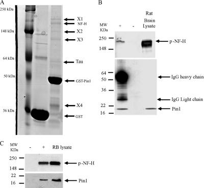
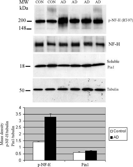
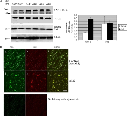
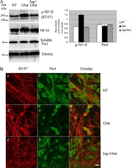
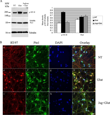
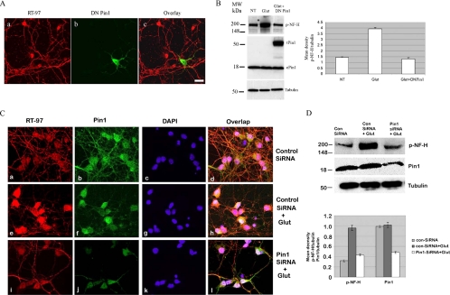
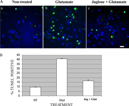
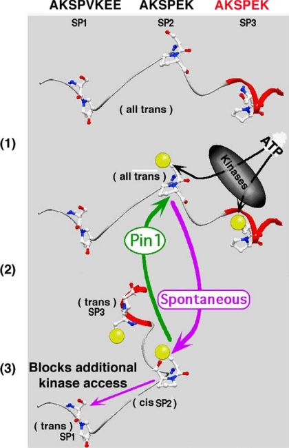
Under normal conditions, the proline-directed serine/threonine residues of neurofilament tail-domain repeats are exclusively phosphorylated in axons. In pathological conditions such as amyotrophic lateral sclerosis (ALS), motor neurons contain abnormal perikaryal accumulations of phosphorylated neurofilament proteins. The precise mechanisms for this compartment-specific phosphorylation of neurofilaments are not completely understood. Although localization of kinases and phosphatases is certainly implicated, another possibility involves Pin1 modulation of phosphorylation of the proline-directed serine/threonine residues. Pin1, a prolyl isomerase, selectively binds to phosphorylated proline-directed serine/threonine residues in target proteins and isomerizes cis isomers to more stable trans configurations. In this study we show that Pin1 associates with phosphorylated neurofilament-H (p-NF-H) in neurons and is colocalized in ALS-affected spinal cord neuronal inclusions. To mimic the pathology of neurodegeneration, we studied glutamate-stressed neurons that displayed increased p-NF-H in perikaryal accumulations that colocalized with Pin1 and led to cell death. Both effects were reduced upon inhibition of Pin1 activity by the use of an inhibitor juglone and down-regulating Pin1 levels through the use of Pin1 small interfering RNA. Thus, isomerization of lys-ser-pro repeat residues that are abundant in NF-H tail domains by Pin1 can regulate NF-H phosphorylation, which suggests that Pin1 inhibition may be an attractive therapeutic target to reduce pathological accumulations of p-NF-H.


Figures

Figure 1.
Figure 1.
Pin1 binds to phosphorylated neurofilament-heavy chain (p-NF-H). (A) GST pulldown assays in rat brain lysates. Samples were separated by 10% SDS-PAGE, and gels were Coomassie stained. Protein bands corresponding to approximately 200 kDa were excised and identified by MS-MS mass spectrometry. This protein was identified as NF-H (arrowed). Other bands present in the gel are as follows: X1, MAP1b; X2, NF-M; X3, keratin; and X4, GST-Pin1 degradation product. Tau, GST-Pin1, and GST are also indicated. A representative gel from five experiments with identical results is shown. (B) A co-IP using anti-Pin1 antibody (Oncogene). Samples were separated by SDS-PAGE and subjected to Western blotting using the RT-97 antibody, which specifically detects p-NF-H. Arrowed band shows the species co-IP with Pin1. + and −, presence and absence of immunoprecipitating antibody, respectively. (C) Co-IP was done using RT-97 antibody to immunoprecipitate p-NF-H. Pin1 was detected in the immunoprecipitate. Arrowed bands shows p-NF-H and Pin1 species. + and −, presence and absence of immunoprecipitating antibody, respectively.
Figure 2.
Figure 2.
NF-H phosphorylation is increased in Alzheimer's disease (AD) tissue. Top panels, total protein lysates from age/region-matched controls (CON) and AD brain were made and separated by SDS-PAGE. The presence of p-NF-H (top panel) and Pin1 (third panel) were immunodetected, and equal loading was confirmed by tubulin. The increase in p-NF-H was evident in AD samples without any change in soluble Pin1 levels. Quantitation is shown in the bar graph below expressed as ±SEM of densitometric measurements of p-NF-H and Pin1 signals normalized to tubulin measurements. Total NF levels were unchanged.
Figure 3.
Figure 3.
NF-H phosphorylation and aggregation are increased in ALS spinal cord tissues. (A) Total lysates of spinal cord tissues from control and ALS were separated by SDS-PAGE. The Western blot shows an increase in phosphorylated NF-H in ALS tissue compared with the controls (CON) without any significant change in Pin1 levels. Quantitation is shown in the bar graph at right, with tubulin showing equal loading. p-NF-H and Pin1 densities were normalized to tubulin densities (obtained through densitometric scanning) expressed as ±SEM. Total NF levels were unchanged. (B) Spinal cord sections from ALS patients and closely matched controls were immunostained for Pin1 (red) and p-NF-H using RT-97 (green). In the ventral region of gray matter the distribution of both Pin1 and p-NF-H were uniform in the area. (g–i) However, in the ventral horn of the white matter of ALS spinal cord, p-NF-H and Pin1 colocalized in aggregates (j–l). Controls without primary antibody did not show any staining. Scale bars, 20 μm.
Figure 4.
Figure 4.
Glutamate excitotoxcity induces p-NF-H accumulations in DRG neuronal cell bodies. (A) Five-day-old DRG neurons were nontreated, treated with 10 mM glutamate for 4 h, or pretreated with 30 μM juglone then treated with 10 μM glutamate for 4 h. Total lysates from the DRG samples were subjected to Western blotting to detect p-NF-H and Pin1. p-NF-H increased upon treatment with glutamate, which was reduced to nontreated levels when DRG neurons were pretreated with juglone before glutamate exposure. Tubulin expression served as loading control. Quantitation shown in bar graph at right as ±SEM of four separate experiments. (B) Five-day-old DRG neurons were nontreated (a–c), treated with 10 μM glutamate for 6 h (d–f) or pretreated with 30 mM juglone and then treated with 10 μM glutamate for 6 h (g–i). In nontreated DRG neurons, Pin1 was localized to the cell body, whereas p-NF-H was found mainly in the processes. On glutamate treatment there was an increase in p-NF-H and colocalization with Pin1 in the cell bodies. This was reduced when neurons were pretreated with juglone before glutamate exposure. Scale bar, 20 μm.
Figure 5.
Figure 5.
Glutamate-mediated increases in p-NF-H levels in cortical neurons is inhibited by juglone. (A) Seven-day-old primary cortical neurons were nontreated, treated with 0.1 mM glutamate for 6 h, or pretreated with 30 μM of the Pin1 inhibitor juglone for 3 h and then treated with 0.1 mM glutamate for 6 h. Total lysates were subjected to Western blotting. p-NF-H increased in the glutamate (Glut)-treated samples and were reduced to nontreated (NT) levels when neurons were treated with juglone before glutamate exposure (Jug+Glut). Soluble Pin1 levels did not change. Bar graph on the right shows the results of four experiments expressed as ±SEM. (B) Neurons were nontreated (NT, a–d), glutamate treated (Glut, e–h), and pretreated with juglone before glutamate exposure (Jug+Glut, i–l), fixed and stained for p-NF-H using RT-97 antibody (red), Pin1 (green), and DAPI (blue). Nontreated neurons exhibited p-NF-H staining in the processes with little or no staining in the cell body, which increased upon glutamate treatment. Cell body p-NF-H staining was reduced when neurons were pretreated with the Pin1 inhibitor before glutamate treatment. Scale bar, 20 μm.
Figure 6.
Figure 6.
Glutamate-mediated increase in phosphorylated NF-H is reduced by overexpression of DN-Pin1 and Pin1-siRNA. (A) Five-day-old cortical neurons were transfected with DN-Pin1 and after 5 h were treated with 0.1 mM glutamate (Glut). Neurons were immunostained and p-NF-H was detected using RT-97 (red) and DN Pin1 detected through expression of GFP. Only neurons transfected with DN Pin1 exhibited reduced phosphorylated NF-H in the cell body. Scale bar, 20 μm. (B) Identical samples from A were harvested for lysates. Total cell lysates were made and separated by SDS-PAGE and then subjected to Western blotting. Phosphorylated NF-H (p-NF-H) was detected using RT-97. Pin1 was immunodetected using anti-Pin1 antibody. p-NF-H was reduced in the DN-Pin1–transfected sample. Transfected Pin1 (tPin1) migrated at approximately 50 kDa because it was a GFP fusion protein, compared with 18 kDa of endogenous Pin1 (ePin1). Equal loading was confirmed by detection of tubulin. The Western blot panels are representative of five independent experiments, and quantitation of p-NF-H is shown in the bar graph on the right, where densitometric measurements of RT-97 were normalized to tubulin measurements and expressed as ±SEM. (C) Five-day-old neurons were transfected with either control siRNA (a–d and e–h) or Pin1-siRNA (i–l) and then treated with 0.1 mM glutamate (Glut, e–h and i–l). Neurons were immunostained, p-NF-H was detected using RT-97 (red), and Pin1 was detected by using Pin1 antibody. Only neurons transfected with Pin1-siRNA exhibited reduced p-NF-H in the cell body. Scale bar, 20 μm. (D) Identical samples from C were harvested and separated by Western blotting for immunodetection of p-NF-H and Pin1. Signals were normalized to tubulin levels. Pin1-siRNA reduced glutamate-induced increases in p-NF-H.
Figure 7.
Figure 7.
Inhibition of Pin1 reduces glutamate mediated neuronal cell death. (A) Seven-day-old cortical neurons were nontreated (a), treated with 0.1 mM glutamate for 6 h (b), or pretreated with juglone then glutamate (c) and presence of apoptotic neurons examined by TUNEL-FITC staining. Nuclei were counterstained using DAPI. TUNEL-positive cells increased upon glutamate treatment and declined after neurons were treated with juglone before glutamate treatment. Scale bar, 20 μm. (B) Quantitation is shown in the bar graph where five independent areas containing a minimum of 50 neurons were counted. Neuronal death increased nearly four times upon exposure to glutamate, and this was reduced nearly twofold when neurons were treated with juglone before glutamate treatment.
Figure 8.
Figure 8.
Hypothetical mechanism for Pin1 regulation of NF-H phosphorylation. The three adjacent KSP repeat units used to illustrate this model are the human NF-H sequence 742–761. We arbitrarily diagram the kinase phosphorylation of NF tail domain repeats as normally starting at the most C-terminal repeat unit and proceeding toward their N-terminus. The more N-terminal repeat units are assumed to be sterically shielded from kinase access by burial within the tail domain until the adjacent C-terminal units are phosphorylated: (1) Tail domain phosphorylation occurs at kinase-accessible C-terminal repeats (yellow spheres); phosphorylation “unwinds” the outer repeat units and permits kinase access to additional repeat units. (2) In some cases, two or more phosphorylations will initiate a trans to cis isomerization of the last phosphorylated S-P bond. This causes a local conformation that does not expose further tail domain phosphorylation sites. However, if active Pin1 is available it will rapidly return the cis p-S/T-P, to trans and allow additional phosphorylation to proceed normally in axons. (3) However, if this occurs in perikarya, premature extension of sidearms may prevent neurofilament subunit transport out of perikarya and cause aggregation of p-NF-H subunits.

Similar articles

Cited by

References

    1. Al-Chalabi A., Miller C. C. Neurofilaments and neurological disease. Bioessays. 2003;25:346–355. - PubMed
    1. Augustinack J. C., Sanders J. L., Tsai L. H., Hyman B. T. Colocalization and fluorescence resonance energy transfer between cdk5 and AT8 suggests a close association in pre-neurofibrillary tangles and neurofibrillary tangles. J. Neuropathol. Exp. Neurol. 2002a;61:557–564. - PubMed
    1. Augustinack J. C., Schneider A., Mandelkow E. M., Hyman B. T. Specific tau phosphorylation sites correlate with severity of neuronal cytopathology in Alzheimer's disease. Acta Neuropathol. 2002b;103:26–35. - PubMed
    1. Bao L., Kimzey A., Sauter G., Sowadski J. M., Lu K. P., Wang D. G. Prevalent overexpression of prolyl isomerase Pin1 in human cancers. Am. J. Pathol. 2004;164:1727–1737. - PMC - PubMed
    1. Becker E. B., Bonni A. Pin1 mediates neural-specific activation of the mitochondrial apoptotic machinery. Neuron. 2006;49:655–662. - PubMed

Publication types

MeSH terms

LinkOut - more resources