. 2007 Sep;18(9):3591-600.
doi: 10.1091/mbc.e07-01-0035. Epub 2007 Jul 11.

Gleevec increases levels of the amyloid precursor protein intracellular domain and of the amyloid-beta degrading enzyme neprilysin

Affiliations

Gleevec increases levels of the amyloid precursor protein intracellular domain and of the amyloid-beta degrading enzyme neprilysin

Yvonne S Eisele et al. Mol Biol Cell. 2007 Sep.

Abstract

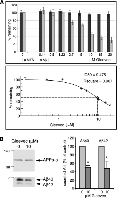
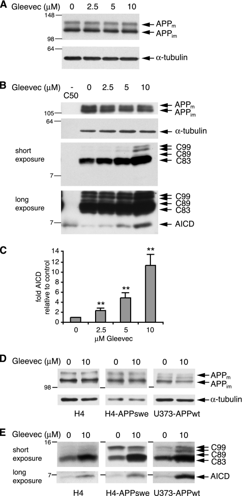
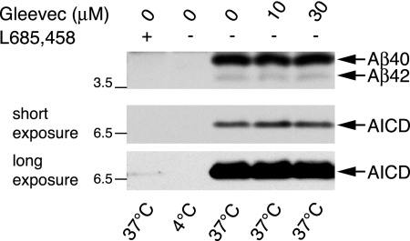
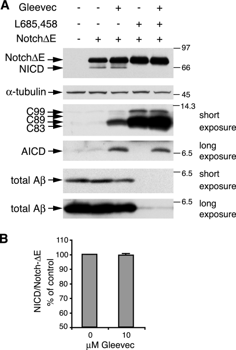
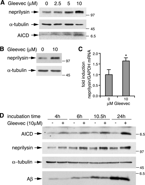
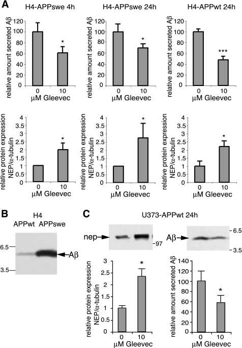
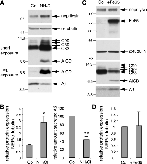
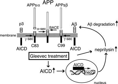
Amyloid-beta (Abeta) deposition is a major pathological hallmark of Alzheimer's disease. Gleevec, a known tyrosine kinase inhibitor, has been shown to lower Abeta secretion, and it is considered a potential basis for novel therapies for Alzheimer's disease. Here, we show that Gleevec decreases Abeta levels without the inhibition of Notch cleavage by a mechanism distinct from gamma-secretase inhibition. Gleevec does not influence gamma-secretase activity in vitro; however, treatment of cell lines leads to a dose-dependent increase in the amyloid precursor protein intracellular domain (AICD), whereas secreted Abeta is decreased. This effect is observed even in presence of a potent gamma-secretase inhibitor, suggesting that Gleevec does not activate AICD generation but instead may slow down AICD turnover. Concomitant with the increase in AICD, Gleevec leads to elevated mRNA and protein levels of the Abeta-degrading enzyme neprilysin, a potential target gene of AICD-regulated transcription. Thus, the Gleevec mediated-increase in neprilysin expression may involve enhanced AICD signaling. The finding that Gleevec elevates neprilysin levels suggests that its Abeta-lowering effect may be caused by increased Abeta-degradation.


Figures

Figure 1.
Figure 1.
Dose-dependent decrease in secreted Aβ but not in secreted APPs-α after Gleevec treatment. (A) Conditioned medium from H4-APPwt cells treated with increasing Gleevec concentrations for 20 h was analyzed for total secreted Aβ by sandwich-ELISA. In parallel, the viability of treated cells was monitored by MTS assay. With increasing Gleevec concentrations, secreted Aβ decreased (top, light gray bars), whereas the viability of cells was unaffected by Gleevec (top, dark gray bars). The IC50 value for inhibition of Aβ secretion was calculated by nonlinear curve fitting of percentage of remaining Aβ values. (B) Analysis of Aβ40 and Aβ42 from conditioned medium of H4-APPwt cells treated with DMSO or 10 μM Gleevec for 24 h. Aβ was immunoprecipitated and analyzed by Western blot by using 6E10 antibody. Band intensities of Aβ40 and Aβ42 were quantified by densitometric analysis, and relative values are shown as percentage of control (n = 4, error bars represent SD; *p < 0.01, unpaired t test). Gleevec reduced secreted Aβ40 and Aβ42 by ∼50%. Levels of secreted APPs-α, as analyzed by Western blot with 6E10 antibody, remained unchanged by Gleevec treatment.
Figure 2.
Figure 2.
Dose-dependent increase in AICD and APP C-terminal fragments after Gleevec treatment. H4-APPwt cells were treated with increasing Gleevec concentrations as indicated for 24 h. (A) Full-length APP from cell lysates was analyzed by 8% SDS-PAGE and Western blot by using 6E10 antibody. Mature (m) and immature (im) forms of APP are indicated. α-tubulin was detected as loading control. (B) AICD and APP C-terminal fragments were separated by 16.5% Tricine SDS-PAGE and detected with antibody A8717. Full-length APP levels remained unchanged, whereas APP C-terminal fragments C83, C89, and C99 (short exposure), as well as AICD (long exposure) showed a Gleevec-dependent increase. Synthetic C50 peptide was loaded as a size control for AICD (see first lane). (C) Densitometric analysis of AICD band intensities. The -fold increase in AICD relative to control is shown (n = 5, error bars represent SD; **p < 0.001, unpaired t test). (D and E) H4, H4-APPswe or U373-APPwt cells were treated with DMSO or 10 μM Gleevec for 24 h. (D) Cell lysates were analyzed for full-length APP as described in A. (E) Analysis of AICD and APP C-terminal fragments as in B. All three cell lines showed a Gleevec dose-dependent increase in AICD and APP C-terminal fragments, whereas APP expression remained unchanged.
Figure 3.
Figure 3.
Gleevec does not directly influence γ-secretase activity in vitro. Generation of AICD and Aβ was analyzed in vitro by using membrane preparations of H4-APPswe cells containing the γ-secretase complex and its substrate C99. Incubation of membrane fractions at 37°C led to the generation of Aβ and AICD by γ-secretase, whereas one reaction was kept at 4°C as a negative control with no enzymatic activity. To test a potential effect on γ-secretase activity in vitro, 10 or 30 μM Gleevec or 1 μM γ-secretase inhibitor L-685,458 was included in the reactions where indicated. After termination of the reactions, Aβ levels were analyzed by Western blot using 6E10 antibody (top) and levels of AICD were detected with antibody A8717 (middle and bottom). Middle, a short exposure to compare Gleevec-treated samples. Bottom, a longer exposure detecting minor amounts of AICD in L-685,458–treated samples but not in the 4°C negative control.
Figure 4.
Figure 4.
Influence of Gleevec and γ-secretase inhibitor L-685,458 on Notch cleavage, APP C-terminal fragments, and Aβ. (A) NICD generation was analyzed by transient transfection of H4-APPswe cells with a myc-tagged NotchΔE construct, which is constitutively cleaved by γ-secretase to generate NICD. Cells were transfected and immediately treated with DMSO, 10 μM Gleevec, 1 μM γ-secretase inhibitor L-685,458, or with both 10 μM Gleevec and 1 μM L685,458 in combination for 24 h. Subsequently, NotchΔE expression and NICD generation were analyzed in cell lysates by using myc-tag–specific antibody 9E10 (top). The amount of APP C-terminal fragments in lysates was assessed by Western blot with antibody A8717 (middle). A shorter exposure to detect C99, C89, and C83 and a longer exposure detecting AICD from the same blot is shown. Total Aβ from conditioned cell media was analyzed with 6E10 antibody (bottom). Two different exposures from the same blot are shown. (B) Band intensities of NICD and NotchΔE were quantified by densitometric analysis from three independent experiments and NICD/NotchΔE ratios were calculated. Gleevec treatment did not influence NICD generation.
Figure 5.
Figure 5.
Gleevec treatment leads to up-regulation of neprilysin protein and mRNA levels. (A) Analysis of neprilysin and AICD levels in Gleevec-treated H4-APPswe cells. After treatment of cells with increasing Gleevec concentrations for 20 h, cell lysates were analyzed by Western blot with the neprilysin-specific antibody 56C6, and subsequently with an α-tubulin specific antibody, serving as a loading control. AICD from cell lysates was analyzed with antibody A8717. Parallel to an increase in AICD up-regulated expression of neprilysin was observed. (B) Analysis of neprilysin protein levels in H4-APPwt cells. Cells were treated with 10 μM Gleevec or DMSO for 15 h and neprilysin and α-tubulin protein levels were analyzed by Western blot as described in A. (C) Analysis of neprilysin mRNA levels from H4-APPwt cells treated as described in B. Neprilysin expression was measured by real-time PCR and normalized to expression of the housekeeping gene GAPDH. The -fold change of neprilysin mRNA in Gleevec-treated cells was 1.64-fold up-regulated (n = 7, error bars represent SD; *p < 0,05, Pair Wise Fixed Reallocation Randomisation Test). (D) Time course of AICD and neprilysin increase and Aβ decrease. H4-APPswe cells, treated with 10 μM Gleevec or DMSO for the times indicated, were lysed and AICD, neprilysin, and α-tubulin were analyzed as described in A. Aβ from conditioned cell media was analyzed by Western blot with 6E10 antibody. As expected, total Aβ in the cell medium increased over time from 4 to 24 h. Comparison of Aβ from Gleevec-treated cells to controls per time point showed a Gleevec mediated reduction in total Aβ.
Figure 6.
Figure 6.
Gleevec-mediated neprilysin up-regulation and concomitant decrease in secreted Aβ in different cell lines. (A) H4-APPswe and H4-APPwt cells treated with 10 μM Gleevec or DMSO for the indicated times were analyzed for secreted Aβ and neprilysin expression by Western blot and quantified by densitometric analysis. Neprilysin expression was normalized to α-tubulin. The relative amount compared with controls is shown. H4-APPswe cells treated with Gleevec for 4 h showed a twofold increase in neprilysin and a decrease in secreted Aβ to 61% of control (n = 8 and n = 3, respectively). In H4-APPswe cells 24 h after Gleevec treatment a 2.7-fold increase in neprilysin and a decrease in secreted Aβ to 70% of control was measured (n = 4 and n = 3, respectively). Neprilysin upregulation in H4-APPwt cells was 2.2-fold, and Aβ secretion was decreased to 48% of control (n = 4). Error bars represent SD; *p < 0.05, ***p < 0.0001, unpaired t test. (B) H4-APPswe cells secrete six- to eightfold higher levels of Aβ than H4-APPwt cells due to the Swedish mutation. Aβ levels from conditioned medium of both cell lines is compared by Western blot analysis. (C) Analysis of neprilysin expression and Aβ secretion in U373-APPwt cells after 24 h of Gleevec treatment. Neprilysin expression normalized to α-tubulin was 2.3-fold, and Aβ secretion was reduced to 57% of controls (n = 3, error bars represent SD; *p < 0.05, unpaired t test).
Figure 7.
Figure 7.
Increase in AICD by alkalizing agent NH4Cl and by Fe65 overexpression have different effects on neprilysin and Aβ. (A) Treatment of H4-APPwt cells with 5 mM NH4Cl led to a strong increase in APP C-terminal fragments, including AICD. Concomitantly, neprilysin protein levels were increased and Aβ levels were decreased. Densitometric analysis of relative neprilysin expression and relative amount of secreted Aβ is shown in B. Neprilysin was 2.8-fold up-regulated, and Aβ secretion was decreased to 43% of control (n = 3, error bars represent SD; *p < 0.01, **p < 0.001, unpaired t test). (C) H4-Fe65i cells, expressing Fe65 under control of the tet-off system, were maintained with doxycycline for 3 d (Co), or Fe65 overexpression was induced by cultivating cells without doxycycline for 3 d (+Fe65). Fe65-expression, neprilysin, APP C-terminal fragments, and Aβ from these cells are shown. Although Fe65 overexpression led to an increase in AICD, larger APP C-terminal fragments were decreased. Concomitantly, Aβ levels increased. Neprilysin expression was not significantly affected by Fe65-mediated AICD induction. Densitometric quantification is shown in D.
Figure 8.
Figure 8.
Working Model of Gleevec Mechanism. Gleevec treatment increases AICD levels via a slowed turnover of AICD. Neprilysin expression is increased by Gleevec, mediated by transcriptional activation, which probably involves AICD signaling. Increased neprilysin expression may lower Aβ levels by enhanced degradation. α-sec, α-secretase; γ-sec, γ-secretase; nep, neprilysin gene.

References

    1. Ando K., Iijima K. I., Elliott J. I., Kirino Y., Suzuki T. Phosphorylation-dependent regulation of the interaction of amyloid precursor protein with Fe65 affects the production of beta-amyloid. J. Biol. Chem. 2001;276:40353–40361. - PubMed
    1. Aplin A. E., Gibb G. M., Jacobsen J. S., Gallo J. M., Anderton B. H. In vitro phosphorylation of the cytoplasmic domain of the amyloid precursor protein by glycogen synthase kinase-3beta. J. Neurochem. 1996;67:699–707. - PubMed
    1. Baek S. H., Ohgi K. A., Rose D. W., Koo E. H., Glass C. K., Rosenfeld M. G. Exchange of N-CoR corepressor and Tip60 coactivator complexes links gene expression by NF-kappaB and beta-amyloid precursor protein. Cell. 2002;110:55–67. - PubMed
    1. Bentahir M., Nyabi O., Verhamme J., Tolia A., Horre K., Wiltfang J., Esselmann H., De Strooper B. Presenilin clinical mutations can affect gamma-secretase activity by different mechanisms. J. Neurochem. 2006;96:732–742. - PubMed
    1. Borg J. P., Ooi J., Levy E., Margolis B. The phosphotyrosine interaction domains of X11 and FE65 bind to distinct sites on the YENPTY motif of amyloid precursor protein. Mol. Cell. Biol. 1996;16:6229–6241. - PMC - PubMed

Publication types

MeSH terms

LinkOut - more resources