. 2007 Sep;18(9):3656-66.
doi: 10.1091/mbc.e07-01-0025. Epub 2007 Jul 11.

Endocytic down-regulation of ErbB2 is stimulated by cleavage of its C-terminus

Affiliations

Endocytic down-regulation of ErbB2 is stimulated by cleavage of its C-terminus

Mads Lerdrup et al. Mol Biol Cell. 2007 Sep.

Abstract

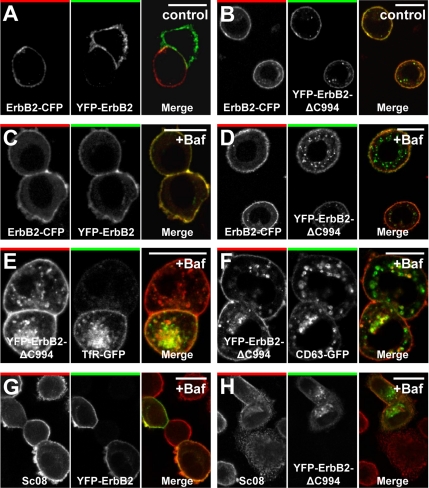
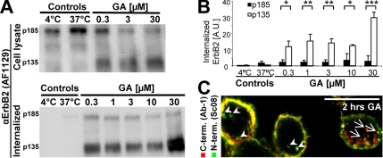
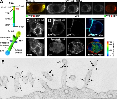
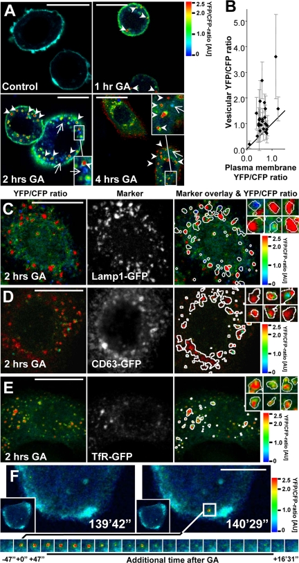
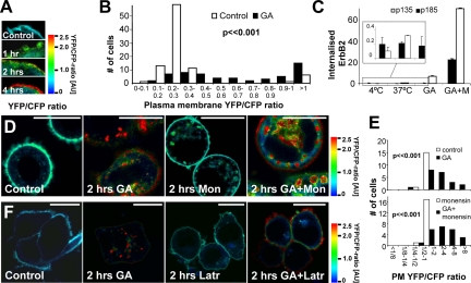
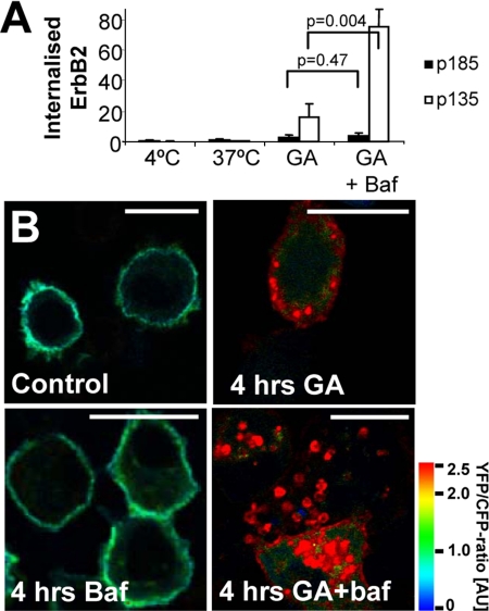
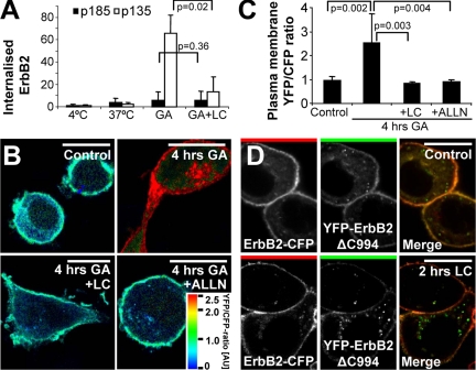
High ErbB2 levels are associated with cancer, and impaired endocytosis of ErbB2 could contribute to its overexpression. Therefore, knowledge about the mechanisms underlying endocytic down-regulation of ErbB2 is warranted. The C-terminus of ErbB2 can be cleaved after various stimuli, and after inhibition of HSP90 with geldanamycin this cleavage is accompanied by proteasome-dependent endocytosis of ErbB2. However, it is unknown whether C-terminal cleavage is linked to endocytosis. To study ErbB2 cleavage and endocytic trafficking, we fused yellow fluorescent protein (YFP) and cyan fluorescent protein (CFP) to the N- and C-terminus of ErbB2, respectively (YFP-ErbB2-CFP). After geldanamycin stimulation YFP-ErbB2-CFP became cleaved in nonapoptotic cells in a proteasome-dependent manner, and a markedly larger relative amount of cleaved YFP-ErbB2-CFP was observed in early endosomes than in the plasma membrane. Furthermore, cleavage took place at the plasma membrane, and cleaved ErbB2 was internalized and degraded far more efficiently than full-length ErbB2. Concordantly, a C-terminally truncated ErbB2 was also readily endocytosed and degraded in lysosomes compared with full-length ErbB2. Altogether, we suggest that geldanamycin leads to C-terminal cleavage of ErbB2, which releases the receptor from a retention mechanism and causes endocytosis and lysosomal degradation of ErbB2.


Figures

Figure 1.
Figure 1.
C-terminally truncated ErbB2 is readily internalized. (A and B) Confocal microscopy images of fixed SK-BR-3 cells transfected with ErbB2-CFP together with YFP-ErbB2 (A) or YFP-ErbB2ΔC994 (B) constructs based on rat ErbB2. (A and B) Left, CFP distribution; middle, YFP distribution; right, the merged signal (CFP, red; YFP, green). In contrast to full-length ErbB2 the truncated version becomes internalized to a detectable level. (C and D) Images similar to the ones in A and B, but cells were incubated with 500 nM bafilomycin for 2 h. Note the increased accumulation of truncated ErbB2-containing endocytic vesicles. (E and F) Confocal images of fixed SK-BR-3 cells transfected with YFP-ErbB2ΔC994 and TfR-GFP (E) or CD63-GFP (F) and incubated with 500 nM bafilomycin for 2 h. Truncated, internalized ErbB2 colocalizes with both markers. (G and H) Confocal microscopy images of fixed, bafilomycin-treated (500 nM) SK-BR-3 cells showing endogenous (human) ErbB2 labeled with Sc08 against ErbB2's N-terminal part together with transfected YFP-ErbB2 (G) or YFP-ErbB2ΔC994 (H). It is seen that endocytosis and vesicular accumulation after bafilomycin treatment is much higher for YFP-ErbB2ΔC994 than for YFP-ErbB2 and that YFP-ErbB2ΔC994 is present in endosomal and lysosomal compartments. Bars, 20 μm.
Figure 2.
Figure 2.
In GA-stimulated cells ErbB2 is preferentially internalized in a cleaved form. (A) Bottom, Western blot from a biotin internalization assay showing internalized full-length and cleaved ErbB2 (p185 and p135, respectively) in SK-BR-3 cells incubated for 2 h with geldanamycin (GA) at the indicated concentrations. Control cells were incubated with the highest DMSO concentration presented to GA-incubated cells. Top, Western blot of full-length and cleaved ErbB2 in the whole cell lysates from which the biotinylated ErbB2 was precipitated. (B) Quantification of internalized ErbB2 measured in arbitrary units (AU) from Western blots like the one in A; n = 3, error bars, SD; *p < 0.05, **p < 0.01, ***p < 0.001, in a two-sided Student's t test. (C) Confocal image of SK-BR-3 cells incubated with 3 μM GA for 2 h and then stained for N- and C-terminal, endogenous ErbB2 (Sc08, green, and Ab-1, red, respectively). Arrows and arrowheads point at structures with predominant C- and N-terminal staining, respectively. Bar, 20 μm.
Figure 3.
Figure 3.
Validation of YEC. (A) Structure of YEC at the DNA and protein levels. This construct is based on rat ErbB2. The scissor indicates the cleavage after the signal sequence (SS). (B) Time-lapse, live cell, confocal microscopy of SK-BR-3 cells transfected with YEC showing the effect of trypsin on YEC. Left image, the localization and intensity of YFP and CFP before incubation with 0.05% trypsin (yellow indicates colocalization). Middle images, the change in YFP intensity (extracellular) only. Right image, the localization and intensity of YFP and CFP after few minutes of trypsin incubation. It is seen that the YFP-tagged, N-terminal part of YEC is indeed extracellular and therefore sensitive to trypsin. (C) Confocal microscopy images showing the localization of YEC (YFP, top) or endogenous ErbB2 (Sc08, bottom) in the same YEC-transfected, fixed SK-BR-3 cell after 2 h of GA incubation. YEC clearly colocalizes with endogenous ErbB2. (D) Live cell confocal microscopy of SK-BR-3 cells transfected with YEC. Left images, the localization of CFP; middle, the localization of YFP; and right, the ratio between YFP and CFP as explained in the color bar. Red, a high YFP-CFP ratio; blue, a low YFP-CFP ratio. Please note that the YFP-CFP ratio is based on arbitrary fluorescence intensities and that a ratio of 1.0 does not correspond to a 1:1 molar ratio between YFP and CFP. The intensity of each pixel in the image is encoded by average YFP and CFP fluorescence. Top, a normal SK-BR-3 cell; bottom, an apoptotic cell. (E) Electron microscopy image of an immunogold-labeled SK-BR-3 cell transfected with YEC. Gold particles, 10-nm, mark YFP (thick arrows); 5-nm gold particles mark endogenous ErbB2 (thin arrows). Bars, (B–D) 20 μm; (E) 500 nm.
Figure 4.
Figure 4.
YEC in endocytic compartments shows a high YFP-CFP ratio. (A) Ratio images of YFP-CFP in YEC expressing SK-BR-3 cells after 0–4 h of 3 μM GA treatment followed by fixation and permeabilization. Arrows and arrowheads point at vesicles with predominant CFP (blue) or YFP (red) staining, respectively. (B) Quantification of plasma membrane versus vesicular YFP-CFP-ratios. Each dot is a cell; error bars, SD. (C–E) Confocal images of fixed SK-BR-3 cells transfected with YEC and GFP-tagged markers for endosomes or lysosomes after 2 h of 3 μM GA treatment. Left images, the YFP-CFP ratio. Middle images, the intracellular marker localization (Lamp1-GFP, CD63-GFP, and TfR-GFP in C–E, respectively). Right images, the ratio images with an overlay of the intracellular marker localization shown as white circumferences. Inserted images, enlargements of YEC and marker positive vesicles. It is seen that Lamp1-GFP, CD63-GFP, and TfR-GFP–containing vesicles generally have a high YFP-CFP ratio. (F) Live cell 4D ratio imaging of a GA-stimulated SK-BR-3 cell. Left image, the cell 139 min and 42 s after incubation with 3 μM GA and just before the formation of a vesicle with high YFP-CFP ratio. Right images, the cell just after formation of the vesicle. The bottom panel of images tracks the fate of the vesicle in the following 16 min and 31 s. It is seen that the vesicle initially has a high YFP-CFP ratio that is reduced after some time because of acidification. Bars, 20 μm.
Figure 5.
Figure 5.
ErbB2 cleavage occurs at the plasma membrane and is not due to recycling from the endocytic pathway. (A) Ratio images of the plasma membrane of fixed, YEC-transfected SK-BR-3 cells treated with 3 μM GA for 0–4 h. (B) Quantitative ratiometry of individual cell membranes from living YEC-transfected SK-BR-3 cells. □, untreated controls; ■, GA-treated cells (2 h). Column height represents the number of cells with the particular ratios. Data were tested with a two-sided Mann-Whitney U test. (C) Biotin internalization assay showing the effect of 10 μM monensin (M) on the 3 μM GA-induced internalized pool of full-length and cleaved ErbB2 after 2 h. Insert, the relatively low values of the first five samples are shown with a scaled-up Y-axis. It is seen that monensin causes a marked increase in intracellular ErbB2 after GA stimulation, and the relative increase for GA+M compared with GA only samples is highest for full-length ErbB2, suggesting that it recycles more efficiently than cleaved ErbB2. Error bars, SD. (D) Ratio images of fixed YEC-transfected SK-BR-3 cells incubated with 3 μM GA and/or 10 μM monensin for 2 h or left as control. Note that YEC is still cleaved at the plasma membrane after GA stimulation when recycling is inhibited with monensin. (E) Distribution of the YFP-CFP ratio on the plasma membrane (PM) of cells treated as in D. Data were tested with two-sided Mann-Whitney U tests. (F) Ratio images of fixed YEC-transfected SK-BR-3 cells incubated with 20 μg/ml latrunculin A and/or 3 μM GA for 2 h or left as control. Note that YEC is still cleaved at the plasma membrane although latrunculin inhibits accumulation of intracellular YEC after GA stimulation. Bars, 20 μm.
Figure 6.
Figure 6.
Cleavage of ErbB2 increases its lysosomal targeting. (A) Biotin internalization assay showing the effect of 500 nM bafilomycin (baf) on the 3 μM GA-induced internalized pool of full-length and cleaved ErbB2 after 2 h. Data were tested with two-sided Student's t tests. Error bars, SD. (B) Ratio images of fixed, YEC-transfected SK-BR-3 cells incubated with 3 μM GA and/or 500 nM bafilomycin for 4 h or left as control. It is seen that much cleaved but little full-length ErbB2 accumulate intracellularly after GA stimulation if lysosomal degradation is inhibited. Bars, 20 μm.
Figure 7.
Figure 7.
GA-induced cleavage of ErbB2 depends on proteasomal activity. (A) Biotin internalization assay showing the effect of 10 μM of the proteasomal inhibitor lactacystin (LC) on the 3 μM GA-induced internalized pool of full-length and cleaved ErbB2 after 2 h. Data were tested with two-sided Student's t tests. Error bars, SD. (B) Ratio images of YEC-transfected, fixed SK-BR-3 cells incubated with 3 μM GA and when indicated also 10 μM lactacystin or 250 μM of the broad protease inhibitor ALLN for 4 h or left as control. (C) Distribution of the YFP-CFP ratio on the plasma membrane of cells treated as in B. Data were tested with two-sided Mann-Whitney U tests. Error bars, 95% confidence intervals. (D) Confocal microscopy images of fixed SK-BR-3 cells cotransfected with ErbB2-CFP and YFP-ErbB2ΔC994 and either treated with 3 μM lactacystin for 2 h or left untreated. It is seen that cleavage and internalization of full-length ErbB2 depends on proteasomal activity and that YFP-ErbB2ΔC994 is internalized even when proteasomal activity is inhibited. Bars, 20 μm.

Similar articles

Cited by

References

    1. Austin C. D., De Maziere A. M., Pisacane P. I., van Dijk S. M., Eigenbrot C., Sliwkowski M. X., Klumperman J., Scheller R. H. Endocytosis and sorting of ErbB2 and the site of action of cancer therapeutics trastuzumab and geldanamycin. Mol. Biol. Cell. 2004;15:5268–5282. - PMC - PubMed
    1. Baulida J., Kraus M. H., Alimandi M., Di Fiore P. P., Carpenter G. All ErbB receptors other than the epidermal growth factor receptor are endocytosis impaired. J. Biol. Chem. 1996;271:5251–5257. - PubMed
    1. Bendtsen J. D., Nielsen H., von H. G., Brunak S. Improved prediction of signal peptides: SignalP 3.0. J. Mol. Biol. 2004;340:783–795. - PubMed
    1. Bucci C., Thomsen P., Nicoziani P., McCarthy J., van Deurs B. Rab7, a key to lysosome biogenesis. Mol. Biol. Cell. 2000;11:467–480. - PMC - PubMed
    1. Burack M. A., Silverman M. A., Banker G. The role of selective transport in neuronal protein sorting. Neuron. 2000;26:465–472. - PubMed

Publication types

MeSH terms