BMPR1a signaling determines numbers of oligodendrocytes and calbindin-expressing interneurons in the cortex
- PMID: 17626200
- PMCID: PMC6672617
- DOI: 10.1523/JNEUROSCI.1434-07.2007
BMPR1a signaling determines numbers of oligodendrocytes and calbindin-expressing interneurons in the cortex
Abstract
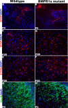
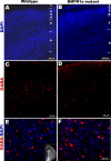
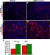
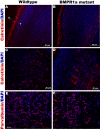
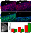
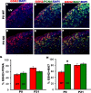
Progenitor cells that express the transcription factor olig1 generate several neural cell types including oligodendrocytes and GABAergic interneurons in the dorsal cortex. The fate of these progenitor cells is regulated by a number of signals including bone morphogenetic proteins (BMPs) secreted in the dorsal forebrain. BMPs signal by binding to heteromeric serine-threonine kinase receptors formed by type I (BMPR1a, BMPR1b, Alk2) and type II (BMPRII) subunits. To determine the specific role of the BMPR1a subunit in lineage commitment by olig1-expressing cells, we used a cre/loxP genetic approach to ablate BMPR1a in these cells while leaving signaling from other subunits intact. There was a reduction in numbers of immature oligodendrocytes in the BMPR1a-null mutant brains at birth. However, by postnatal day 20, the BMPR1a-null mice had a significant increase in the number of mature and immature oligodendrocytes compared with wild-type littermates. There was also an increase in the proportion of calbindin-positive interneurons in the dorsomedial cortex of BMPR1a-null mice at birth without any change in the number of parvalbumin- or calretinin-positive cells. These effects were attributable, at least in part, to a decrease in the length of the cell cycle in subventricular zone progenitor cells. Thus, our findings indicate that BMPR1a mediates the suppressive effects of BMP signaling on oligodendrocyte lineage commitment and on the specification of calbindin-positive interneurons in the dorsomedial cortex.
Figures




Similar articles
-
Differential modulation of BMP signaling promotes the elaboration of cerebral cortical GABAergic neurons or oligodendrocytes from a common sonic hedgehog-responsive ventral forebrain progenitor species.Proc Natl Acad Sci U S A. 2002 Dec 10;99(25):16273-8. doi: 10.1073/pnas.232586699. Epub 2002 Dec 2. Proc Natl Acad Sci U S A. 2002. PMID: 12461181 Free PMC article.
-
A subpopulation of olfactory bulb GABAergic interneurons is derived from Emx1- and Dlx5/6-expressing progenitors.J Neurosci. 2007 Jun 27;27(26):6878-91. doi: 10.1523/JNEUROSCI.0254-07.2007. J Neurosci. 2007. PMID: 17596436 Free PMC article.
-
BmpR1A is a major type 1 BMP receptor for BMP-Smad signaling during skull development.Dev Biol. 2017 Sep 1;429(1):260-270. doi: 10.1016/j.ydbio.2017.06.020. Epub 2017 Jun 19. Dev Biol. 2017. PMID: 28641928 Free PMC article.
-
The cerebral cortex is a substrate of multiple interactions between GABAergic interneurons and oligodendrocyte lineage cells.Neurosci Lett. 2020 Jan 10;715:134615. doi: 10.1016/j.neulet.2019.134615. Epub 2019 Nov 9. Neurosci Lett. 2020. PMID: 31711979 Review.
-
Cortical interneuron fate determination: diverse sources for distinct subtypes?Cereb Cortex. 2003 Jun;13(6):670-6. doi: 10.1093/cercor/13.6.670. Cereb Cortex. 2003. PMID: 12764043 Review.
Cited by
-
The dynamic role of bone morphogenetic proteins in neural stem cell fate and maturation.Dev Neurobiol. 2012 Jul;72(7):1068-84. doi: 10.1002/dneu.22022. Dev Neurobiol. 2012. PMID: 22489086 Free PMC article. Review.
-
Regulation of GNRH production by estrogen and bone morphogenetic proteins in GT1-7 hypothalamic cells.J Endocrinol. 2009 Oct;203(1):87-97. doi: 10.1677/JOE-09-0065. Epub 2009 Jul 27. J Endocrinol. 2009. PMID: 19635757 Free PMC article.
-
NMDA receptor signaling in oligodendrocyte progenitors is not required for oligodendrogenesis and myelination.J Neurosci. 2011 Aug 31;31(35):12650-62. doi: 10.1523/JNEUROSCI.2455-11.2011. J Neurosci. 2011. PMID: 21880926 Free PMC article.
-
Noggin protects against ischemic brain injury in rodents.Stroke. 2010 Feb;41(2):357-62. doi: 10.1161/STROKEAHA.109.565523. Epub 2009 Dec 17. Stroke. 2010. PMID: 20019326 Free PMC article.
-
The bone morphogenetic protein antagonist noggin protects white matter after perinatal hypoxia-ischemia.Neurobiol Dis. 2011 Jun;42(3):318-26. doi: 10.1016/j.nbd.2011.01.023. Epub 2011 Feb 17. Neurobiol Dis. 2011. PMID: 21310236 Free PMC article.
References
-
- Ahn K, Mishina Y, Hanks MC, Behringer RR, Crenshaw EB., III BMPR-IA signaling is required for the formation of the apical ectodermal ridge and dorsal-ventral patterning of the limb. Development. 2001;128:4449–4461. - PubMed
-
- Alcantara S, Ferrer I, Soriano E. Postnatal development of parvalbumin and calbindin D28K immunoreactivities in the cerebral cortex of the rat. Anat Embryol (Berl) 1993;188:63–73. - PubMed
-
- Alcantara S, de Lecea L, Del Rio JA, Ferrer I, Soriano E. Transient colocalization of parvalbumin and calbindin D28k in the postnatal cerebral cortex: evidence for a phenotypic shift in developing nonpyramidal neurons. Eur J Neurosci. 1996;8:1329–1339. - PubMed
-
- Anderson SA, Qiu M, Bulfone A, Eisenstat DD, Meneses J, Pedersen R, Rubenstein JL. Mutations of the homeobox genes Dlx-1 and Dlx-2 disrupt the striatal subventricular zone and differentiation of late born striatal neurons. Neuron. 1997;19:27–37. - PubMed
-
- Anderson SA, Marin O, Horn C, Jennings K, Rubenstein JL. Distinct cortical migrations from the medial and lateral ganglionic eminences. Development. 2001;128:353–363. - PubMed
MeSH terms
Substances
LinkOut - more resources
Full Text Sources
Molecular Biology Databases
